Page 1 of 2

Patterns in MS Lesions

Posted: Thu Mar 05, 2009 1:30 pm
by cheerleader
In researching why some people respond to steroids, and others do not, I've come across research by Drs. Lucchinetti/Lassmann/Bruck on the Subtypes of Multiple Sclerosis Lesions. These doctors have studied MS lesions for many years, in vivo and on autopsy, and have catagorized lesions into four distinct patterns:

Pattern I
-Centered around venule with sharp, demarcated edges
-T-cell/macrophage associated
-extensive remyelination
-oliogodendrocytes in lesion center

Pattern II
-Antibody/complement associated, lesions contain large quantities of immunoglobulin proteins
-Centered around venule, with sharp demarcated edges
-Deposition of immunoglobins and activated compliment at site of demyelination
-Resembles an antibody mediated process
-No defects in mitochondrail respiratory chain detected
-These lesions respond to plasma exchange, but not steroids....

Pattern III
-Distal oligodendrogliopathy, diffuse lesions with variable inflammation and pronounced microglial activations
-Indistinct border
-not centered around venule
-striking loss of myelin associated glycoprotein
-no compliment activation
-Pattern associated with hypoxia
-dying back of oligodendrocyte
-Looks like white matter stroke
-Represents the early stage of lesions during an exacerbation
-Defects in mitochondrial respitory chain


Pattern IV
-sharp macrophage borders
-More rare, represents PPMS lesions
-Degeneration and oligodendrocyte death in white matter
-inactive plaque and no remyelination
“Multiple active plaques in individual brain autopsies [27 autopsies, 170 lesions] revealed identical morphological and immunopathological alterations,” Dr. Lucchinetti explained. “Patterns 1 and 2 suggest that myelin is the target, while patterns 3 and 4 suggest the oligodendrocyte may be the target. In pattern 1, macrophages likely mediate demyelination, whereas in pattern 2, antibody and complement may contribute to demyelination.” She explained that patterns 1 and 2 resemble autoimmune models of multiple sclerosis, while patterns 3 and 4 resemble viral, toxic, ischemic, or metabolic models.
I found this research fascinating, and didn't remember seeing it discussed on here-so I put the pattern lists together from a variety of research-
For further reading:
link
link
thoughts?
AC
Posted: Thu Mar 05, 2009 2:27 pm
by LR1234
I find this really intereting, and it is probably the reason like Peta said why some people respond to some things and others don't.

If someone responds to steroids does that mean that they are always going to respond (well, until their body becomes immune to it) or if someone hasn't responded to steroids in the past can they suddenly respond in a different relapse?

(that sounds confusing when I write that down but do you get what I mean?!!)

Posted: Thu Mar 05, 2009 3:48 pm
by jimmylegs
good compilation cheer, points up that 'ms' might be a bit of a grab bag for a few diff conditions, doesn't it.

Posted: Thu Mar 05, 2009 4:03 pm
by dignan
Cheer, it's good that you're bringing this forward again. There have been numerous discussions of Lucchinetti's work here, but not for a while, so it's good to inform people who weren't around for the earlier discussions. A good one can be found here:

http://www.thisisms.com/ftopic-330-days ... asc-0.html

Good old Felly. Anybody know what became of Felly?

Posted: Thu Mar 05, 2009 4:09 pm
by Hub
Here's an article that cites Lassmann's classification of 4 types of lesions and also cites other researchers that dispute the validity of the classification.

http://www.jhasim.com/files/articlefile ... tanian.pdf

Posted: Thu Mar 05, 2009 4:15 pm
by mrhodes40
:D I was posting the same link Dignan! I agree that was a memorable discussion and really worth revisiting, I have thought of it often.

I have wondered though if it is the person's genes that result in the lesion heterogeneity vs the cause being heterogenous. :?

Nine years old that research is. It sure has not panned out in a ton of new findings as I thought it would.......... :roll:

When it came out I could still jog.

Posted: Thu Mar 05, 2009 4:28 pm
by mrhodes40
HUb, thanks a bunch for that link! GREAT paper! :D

Posted: Thu Mar 05, 2009 7:49 pm
by cheerleader
Thanks for the links, guys. I didn't pull it up on the boards with the search function, since I was searching Lassmann. Man, it's a shame Finn took off with all of his posts.

Lovely Dr. Luchinetti is still at it, working as director of The MS Lesion Project.
The MS Lesion Project News-Making Excellent Progress

One of their first findings showed that individuals with a specific lesion pattern responded to plasma exchange therapy, a treatment used occasionally to treat individuals experiencing severe MS attacks that do not respond to standard steroid therapy.
The Lesion Project team collaborated with Harvard investigators in a recent study that used novel technology to screen the immune response in blood samples from people with various courses of MS. They were able to differentiate between different types of MS and MS patterns of damage, based on distinct immune antibody signatures against different immune targets. The results could ultimately lead to laboratory tests that help diagnose MS and predict its course. Read details of this study.
Recently the team found evidence that natural myelin repair occurs in patients across most types and stages of MS. Further research should determine why some individuals show efficient myelin repair while others do not, and offer clues to ways to stimulate natural repair. Read details of this study.
In October 2007, the team reported on an analysis of MRI scans on 168 individuals from whom biopsy materials had been collected. Some key findings:
Larger biopsied lesion size was associated with a slightly higher disability score (using the EDSS scale of disability) at last clinical follow-up.
They confirmed an earlier finding that people with either of two lesion patterns often showed ring-shaped patterns on MRI.
Brain tissue samples are essential to efforts such as this. Read more about how to participate in such research.
link

But I agree with you all, I think this lesion study is just old news , now that 7Tesla MRIs are showing teeny tiny lesions on veins....
In our two RRMS patients, we were able to demonstrate a total of 80 MS lesions, 58 and 22 lesions independently, and all lesions showed a strict perivascular distribution, following the form, orientation, and course of the vessels, this feature being best noted in small lesions (Figure 1). The diameter of veins associated with lesions ranged from 0.3mm–0.7mm.
http://www.pubmedcentral.nih.gov/articl ... id=2579786

AC

Posted: Thu Mar 05, 2009 8:13 pm
by rainer
Don't know if this a new link but it includes a 2 page article on the 7T and MS.

4 page pdf:

http://communications.med.nyu.edu/files ... _of_ms.pdf

Posted: Sat Mar 07, 2009 3:30 am
by Wonderfulworld
Interesting, could explain why I have never responded to steroids at all, and why I had some many lesions at diagnosis.

Posted: Tue Mar 24, 2009 1:16 pm
by Punchy
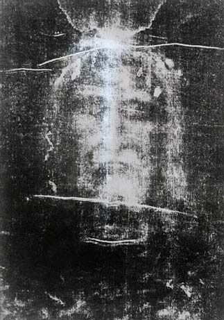
My lesions form a pattern of Jesus' face. I wonder what the significance of that is? 8)

Posted: Tue Mar 24, 2009 1:34 pm
by Lyon
.

Posted: Tue Mar 24, 2009 3:04 pm
by cheerleader
Punchy...
this your MRI?

Image

Posted: Tue Mar 24, 2009 11:07 pm
by marcstck
I've discussed the findings of The Lesion Project with a number of neurologists, and most of them are a bit skeptical of the findings. They say that even if a patient displays only one type of lesion postmortem, that does not mean that their lesions didn't evolve through different stages over time.

I find it striking, though, that the PPMS patients studied all were afflicted with the same type of lesion, and that that lesion type did not show up in any other flavor of MS.

It almost seems like most mainstream neurologists have some kind of vested interest in looking at this disease as a single entity, when more and more evidence shows that multiple sclerosis is more a syndrome and disease.

The latest I've heard of The Lesion Project is that they are actively seeking ways to distinguish lesion type by MRI study. If they could achieve that, it would be a significant breakthrough...