Page 2 of 3

Posted: Sat Oct 17, 2009 3:08 pm
by jimmylegs
think so, LA. hyperintensity. here are a couple of mine:
http://i285.photobucket.com/albums/ll49 ... 1232649925
on the left, my original scan. i am days away from being given the msdx.
on the right, post major dietary mods and vitamin-mania.
the scans are not in exactly the same spot but from what you can see, to me it does look like things improved.

Posted: Sat Oct 17, 2009 3:35 pm
by Absentee
just remember, they cannot correlate "lesions" to symptoms or any of what you are feeling. So try not to get too worked up over what you see.

(typed while prepping my Copaxone shot!)

Posted: Sat Oct 17, 2009 3:39 pm
by LoveActually
Thanks JL!

And thank you Absentee. Prepping a shot to give to myself, I don't think I can do it.

Posted: Sat Oct 17, 2009 3:44 pm
by jimmylegs
correction: when it's spinal lesions, you can. especially when they're from chronic b12 deficiency (which is the main differential dx for ms) from what i recall, it's the brain ones that don't necessarily correlate.

Posted: Sat Oct 17, 2009 6:15 pm
by LoveActually
JL,

Thank you for sharing your images, a few of mine are below. I had almost 600 in total but pulled out some that showed a lot more spots? then others.

I have no idea what I'm looking at but to me, it doesn't look good.

http://tinyurl.com/yfm6pyz

Posted: Sat Oct 17, 2009 6:41 pm
by jimmylegs
LA, i don't have a snapfish account so i can't see. just so you know though, the classic pattern for spinal lesions in b12 deficiency are cervical and thoracic lesions. like mine.

Posted: Sat Oct 17, 2009 7:38 pm
by LoveActually
Sorry about that. Let me see how big they are when I post them here...

Image

Image

Hopefully that's not too large, if so I can remove them. So can all the lesions on my spine (guessing those white spots are lesions) be just from B12 deficiency? By the way, there are 3 on my brain.

Re: Unfortunately, I'm Back

Posted: Sat Oct 17, 2009 8:31 pm
by NHE
LoveActually wrote:Hopefully that's not too large, if so I can remove them. So can all the lesions on my spine (guessing those white spots are lesions) be just from B12 deficiency? By the way, there are 3 on my brain.
They're not a problem. Images posted in the forums can be up to 500 pixels wide without disrupting the formatting of the forum's columns. You could probably go even a little bigger, perhaps up to 700 pixels.

NHE

Posted: Sat Oct 17, 2009 8:37 pm
by LoveActually
Great! Thank you NHE. :D

Posted: Sun Oct 18, 2009 5:14 pm
by jimmylegs
hi LA, yes your spinal lesions can be pure b12 deficiency.

if you only have three lesions on your brain, from what i understand that is not quite enough for them to definitely say ms, if your docs are anything like my neuro. i think he wanted to see at least 9 brain lesions or something like that? does your brain scan write up say anything about 'dawson's fingers'? i have not yet seen those associated in research with anything but ms. and i have tried to find them studied in some other illness, but no luck so far.

they find brain lesions sometimes in people upon autopsy, who never had any symptoms at all so the brain lesion thing is a bit of an iffy diagnostic measure anyway.

if they find O-bands in your spinal fluid, that's a marker for chronic inflammation and they'll say that's not caused by b12 deficiency (i don't actually believe that is true - b12 is anti-inflammatory) and if your case is like mine, that will be the last straw, and they will dx ms.

when i started out at tims i was fixated on b12 because it really was all i knew about as a kind of problem area for strict vegetarians, which i was at the time. so in early 2006 i dug around for studies that would show that my problems could be b12, period. in fact i did find studies where brain lesions and o-bands were seen in b12 deficiency.

of course, i had yet to learn about my d3 deficiency, my zinc deficiency, my sub-optimal uric acid, low magnesium, and all the rest of it 7:P !!!

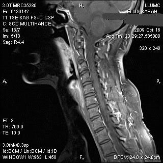
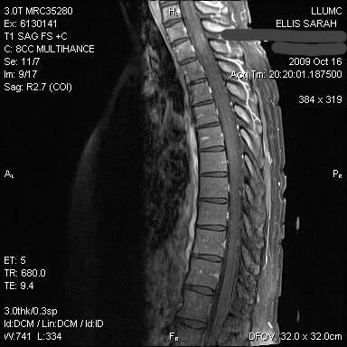
as for where the lesions are, what does it say in the write-up? i'm kind of looking at what i think is the area adjacent to C7, does it mention C7 at all in the mri report? i can't tell which vertebra is which in the thoracic scan, but i think the area about the 3rd v from the top, and the 7/8th from the top look a bit washed out.

the weirdest thing to me is that you have like NO thecal sac. you're supposed to have a protective layer of fluid around your cord, which is the white outline around the darker cord, that you can see on my scan. do you have any other scans maybe, where you can see that white outline?

where my cervical lesion was worst, my cord looked kind of spread out and the sac was thinner, and that's where it looks most like yours, on the left side at least. in my second scan you can see that the cord kind of looks more put together, and the thecal sac appears to be in better shape.

anyway, enough blabbing for now :)
JL

Posted: Sun Oct 18, 2009 6:58 pm
by LoveActually
Hey JL,

The 3 spots on the brain, nothing was mentioned about Dawson's Fingers. My new neuro requested these other tests (spinal MRI, EEG & VEP) because he wasn't comfortable diagnosing on the 3 brain lesions and symptoms alone. He said that judging from the larger spot on my brain and my symptoms, that it's likely I do have MS but he wanted to run more tests to be sure. He said the smaller 2 lesions on my brain could be from migraines but the larger one was definitely not caused by migraines.

I do have other pics where you can see the thecal sac but I haven't pulled them off the CD yet. I don't have the report yet, ye of little patience I am. I should get it on Wednesday.
i can't tell which vertebra is which in the thoracic scan, but i think the area about the 3rd v from the top, and the 7/8th from the top look a bit washed out.
I was looking at that part too. So the white marks on the vertebra are nothing to worry about? I have a lot of those marks and at first I was freaking out. So the washed out part that you were refering to, is that where the lesions would be? Is that the brain stem? Sorry for all the questions, wish I would've paid more attention in school. :)

Has anyone ever heard of someone emailing a Radiologist Report? I live about 40 minutes from the hospital and don't want to make the drive if I don't have to. I'm thinking that may be a privacy problem though. Just curious if anyone was successful with requesting it to be emailed.

Posted: Sun Oct 18, 2009 8:30 pm
by jimmylegs
Hey JL,
The 3 spots on the brain, nothing was mentioned about Dawson's Fingers.
well, that's good!
neuro requested these other tests (spinal MRI, EEG & VEP) because he wasn't comfortable diagnosing on the 3 brain lesions and symptoms alone.
did he mention lumbar puncture? you haven't had that done yet have you?
the smaller 2 lesions on my brain could be from migraines but the larger one was definitely not caused by migraines.
where was the larger one?
So the white marks on the vertebra are nothing to worry about? I have a lot of those marks and at first I was freaking out.
i am not sure about why you might have white marks right on the vertebrae - i have not seen a lot of MRI scans but there is nothing like that on mine, and i don't know what might cause them...
So the washed out part that you were refering to, is that where the lesions would be?
yes i think so.
Is that the brain stem?
no the brain stem is continuous with the spinal cord, but is above the first cervical vertebra (i think)!
Has anyone ever heard of someone emailing a Radiologist Report? I live about 40 minutes from the hospital and don't want to make the drive if I don't have to. I'm thinking that may be a privacy problem though. Just curious if anyone was successful with requesting it to be emailed.
y text report came as an image on the CD with all the MRI scans, so i can't contribute re: emailing, sorry :(

Posted: Mon Oct 19, 2009 7:06 am
by patientx
LoveActually wrote:Has anyone ever heard of someone emailing a Radiologist Report? I live about 40 minutes from the hospital and don't want to make the drive if I don't have to. I'm thinking that may be a privacy problem though. Just curious if anyone was successful with requesting it to be emailed.
I just had one mailed to my home address - but not emailed. I don't know if privacy rules vary by state, but I don't see why they wouldn't mail it to you after verifying it's you who is calling.

My advice would be to take your films to your next neurology appointment, and have the neuro go over them with you. I remember looking the films after my first MRI, and I couldn't make any sense of them. When the neuro started pointing things out to me, I had the "aha" feeling. Plus, things I thought were insignificant were actually lesions, and spots I thought were lesions were nothing (at least according to him). Also, remember the MRI is not a camera - it's really an indirect way of looking at internal tissue, and involves a bunch of computer processing. As such, it is prone to resolution issues and artifacts.

Posted: Mon Oct 19, 2009 8:26 am
by LoveActually
patientx,

Thank you! I always have my 'moments of insanity' after a test and spend hours on the computer trying to figure things out for myself. Now, 3 days after the test, I'm starting to chill out. I'll definitely take the films with me and see what my neuro has to say about them.

JL,

I haven't had it done yet and yes, my neuro did mention a lumbar puncture. He said if all these results come back and he's still on the fence about diagnosing me, he'll request a lumbar puncture. That's one referral I won't mind waiting on. Having that done scares the crap out of me. I've heard all the warnings about the headaches and I'll be sure to stay lying down for as long as possible.

The larger spot is adjacent to the anterior aspect of the left lateral ventricle. That means nothing to be but does it make sense to you?

I just got off the phone with the neurodiagnostic lab and was able to schedule my EEG and VEP for this Wednesday. That's great because they usually schedule 2-3 weeks out but they were able to squeeze me in so I could have everything done by the 29th, my next appt with my neuro. I guess this takes care of the radiologist report issue since I'll be in the area.

This EEG is sleep deprived and will only last about 45 minutes according to the nurse. Much better then walking around with that thing for 72hrs. With the EEG and VEP, can I request a copy of the results at that time or does it take a while to process? Like many of you have advised, I'm trying to get copies of all results.

Posted: Mon Oct 19, 2009 3:52 pm
by jimmylegs
heya LA, yea the LP is no fun that's for sure. as for the larger spot, i know *where* that is but i don't know what the brain matter there does, so without spending a LOT more time on it, i'm not really any help on this one, sorry :(

i hope this works, 'coz it's cool:
Image