Page 329 of 557
Posted: Fri Apr 22, 2011 8:05 pm
by Cece
those are big reasons too, bestadmom, when I thought my list was complete!

Double-checked on Haskal and here is a patient saying Haskal used IVUS on him:
www.thisisms.com/ftopicp-155498.html#155498
Dr. Sclafani's novel use of it to measure the stenosis to match the balloon size is unique to him, as far as I know, but I am encouraged that these other doctors have seen value in IVUS as well.
I wonder if we might get to see venogram vs ivus comparison images of any of the venograms last week (3 out of 9!) in which a stenosis was not seen on venogram but caught on IVUS? I was surprised at the mention that they were valvular stenoses. Those are big and not likely to be missed on the venogram, I would have thought.
Posted: Fri Apr 22, 2011 8:40 pm
by dan46
Dr Cumming used ivus on me. He posted images on the dr to dr thread. There was no reply. I'll give it a bump. You an see them there if you like. I'm the 64 yr. old
dan46
Posted: Fri Apr 22, 2011 11:00 pm
by drsclafani
Cece wrote:drsclafani wrote:i havent noticed anyone speak about it.
That azygous is a real challenge. We have talked about it before.
i used to do 0 degrees, 20 degrees, 70 degrees, 90 degrees until i had a decent understanding of which views offered the most information.
My standard routine currently is to perform an inhalation breathhold, AP view to look at the ascending portion of the azygous vein. I do an upper and a lower view trying to see the entire azygous, hemiazygous and connections to the renal vein. Then I do a 70 degree oblique. With a 70 degree oblique, i do not h ave to raise the arms above the head. It is good in about 90% of cases in seeing the entire arch of the azygous and giving a second view of the ascending azygous. If either the anterior or posterior arch is not well visualized or obscured by superimposition, I will do a 90 degree view. that usually suffices. Then I do IVUS because I do not trust the venogram 100% in most cases.
I love the images, it is something to see how the azygous looks ok in the first view and then the stenosis is seen in the "side view."
Cece
this is a case where the azygous venogram showed reflux but NOTHING in two views to sugest the cause. . However IVUS easily showed a grossly abnormal valve posterior to the arch.
Have a look:
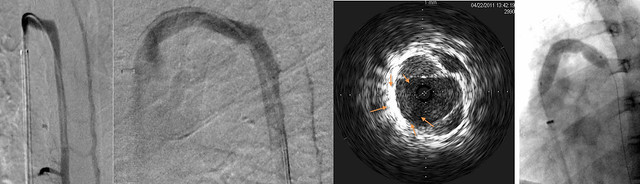
There is a frontal and 70 degree oblique view.The image of IVUS shows thickened valves, outlined with orange arrows . These valves during the movie do not move. the final image on the right shows the waist of the balloon at the valvular obstruction.
Posted: Fri Apr 22, 2011 11:03 pm
by drsclafani
Cece wrote:
We've heard recently that another CCSVI doctor has been finding and treating renal vein obstructions too. What do you think of this? Have you seen any renal vein obstructions and have you looked?
I have found one renal vein stenosis in a patient with azygous disease. the most common finding is hypoplasia of the lumbar veins.
Posted: Fri Apr 22, 2011 11:37 pm
by NZer1
Hi Dr, is disease of the veins a common problem?
If I understand it correctly the people who have benefit from diet changes are likely to be people with disease issues rather that malformations (although the two together would also be possible and likely).
Trying to get my head around the improvement from diet as well.
What is the estimated number of times that IVIS has found issues that would have gone unnoticed?
It seems that the cost issue is outweighed by the outcome!
Or could it be that the issues that IVIS detects are not likely to be problematic in MS?
Keep up the stunning work Dr and thank you so much,
Nigel
Posted: Sat Apr 23, 2011 6:27 am
by Cece
You don't look a day over 46, dan46....
Here's a direct link:
www.thisisms.com/ftopicp-156435.html#156435
I hadn't noticed before that he labelled these ivus pictures. 'Below kink' is narrowed. 'Above kink' is clear. 'At kink' ?
Posted: Sat Apr 23, 2011 6:44 am
by Cece
drsclafani wrote:
Cece
this is a case where the azygous venogram showed reflux but NOTHING in two views to sugest the cause. . However IVUS easily showed a grossly abnormal valve posterior to the arch.
Have a look:
There is a frontal and 70 degree oblique view.The image of IVUS shows thickened valves, outlined with orange arrows . These valves during the movie do not move. the final image on the right shows the waist of the balloon at the valvular obstruction.
I am glad for the orange arrows, because even with them I am having trouble finding the valve. What's fascinating is that you are right, it is hard to see anything in the venogram to indicate it even now that we know it is there.
Posted: Sat Apr 23, 2011 7:05 pm
by lucky125
bestadmom wrote:To add to CeCe's spot-on explanation of why IVUS isn't widely used, currently it is not reimburseable thru insurance. When Dr. S uses it, AAC pays for it, not the patient. It's at least a $600 catheter expense. Then there's the time involved to examine the veins two ways, and the equipment to view the images.
My IVUS was covered by insurance all three times I was treated. Maybe because it was used in a hospital setting, or maybe because the staff at hospitals know how to code it just right. Just FYI!
Posted: Sat Apr 23, 2011 11:54 pm
by drsclafani
NZer1 wrote:Hi Dr, is disease of the veins a common problem?
If I understand it correctly the people who have benefit from diet changes are likely to be people with disease issues rather that malformations (although the two together would also be possible and likely).
Trying to get my head around the improvement from diet as well.
What is the estimated number of times that IVIS has found issues that would have gone unnoticed?
It seems that the cost issue is outweighed by the outcome!
Or could it be that the issues that IVIS detects are not likely to be problematic in MS?
Keep up the stunning work Dr and thank you so much,
Nigel
10%. but the value is in more than detecting lesions. High accuracy in determining diameter is very important too.
Posted: Sat Apr 23, 2011 11:57 pm
by drsclafani
Cece wrote:drsclafani wrote:
Cece
this is a case where the azygous venogram showed reflux but NOTHING in two views to sugest the cause. . However IVUS easily showed a grossly abnormal valve posterior to the arch.
Have a look:
There is a frontal and 70 degree oblique view.The image of IVUS shows thickened valves, outlined with orange arrows . These valves during the movie do not move. the final image on the right shows the waist of the balloon at the valvular obstruction.
I am glad for the orange arrows, because even with them I am having trouble finding the valve. What's fascinating is that you are right, it is hard to see anything in the venogram to indicate it even now that we know it is there.
cece
the arrows are a bit off. There are two diagnonal white lines in the ivus image. they are the valve opening maximally. beyond these lines is the circumferenctial bright signal of the wall of the vein. those valves should open all the way.
Posted: Sun Apr 24, 2011 7:00 am
by Cece
ok, the white lines were what I saw as the edges of the valves until I got confused by the orange arrows.
I hope I am not the only one interested in this here!
Posted: Sun Apr 24, 2011 9:39 am
by 1eye
I will not even attempt to guess what is pointed to by any of the arrows. However, this seems very important:
it is hard to see anything in the venogram to indicate it even now that we know it is there.
This means the IVUS is an indispensable tool without which serious problems
will be missed. It
must be a standard of care, regardless of expense. The expense will grow less, but if there is no other way to find the problems, damn the torpedos!
I MEAN IT! ARE YOU LISTENING?
As a BTW, is IVUS Doppler or just normal?
Posted: Sun Apr 24, 2011 8:09 pm
by drsclafani
1eye wrote:I will not even attempt to guess what is pointed to by any of the arrows. However, this seems very important:
I will try again to illustrate the findings that IVUS detected.
here is the images as before.
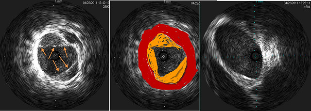
on left is IVUS of malformed immobile valve. Arrows point to the edges of thickened valve tissue. You will have to take my word that those valves do not open completely
in the center is an illustration of the stenosis. The wall of the vein is seen as red. The valve is seen as orange. You can see the stenosis pretty well, even though you cannot see the maximum narrowing of the vein on this image.
on the right is a normal ivus
Posted: Sun Apr 24, 2011 8:18 pm
by drsclafani
1eye wrote:
This means the IVUS is an indispensable tool without which serious problems will be missed. It must be a standard of care, regardless of expense. The expense will grow less, but if there is no other way to find the problems, damn the torpedos! I MEAN IT! ARE YOU LISTENING?
As a BTW, is IVUS Doppler or just normal?
1eye: we cannot make such claims based upon the few anecdotes. This is a substantial expense and we must prove its value.
The most important test would be to determine whether patients have a better outcome when IVUS is used. That is a five year study of durability and clinical outcome.
A less valuable test could be to do an "intent to treat" study. have a treatment plan based upon venography. then reveal the IVUS to the IR being studied (i will bet you guys like that one) to see whether the treatment plan changes.
Of course, we can documenet what percentage of vessels have something uncovered by IVUs, not uncovered by venography. By the way, there are other cases where ivus does not show what venography does.
Lots to study, eh
Posted: Mon Apr 25, 2011 12:23 am
by Johnson
drsclafani wrote:...
Lots to study, eh
Brush up, Doc. I'm on my way to see you. (no pressure, of course. 32psi)
I like your illustration, BTW. Very Miro. (don't forget to clean your brushes.)