Page 375 of 557

Posted: Tue Aug 09, 2011 7:55 pm
by Cece
HappyPoet wrote:I wonder if this woman is the patient who needed to come back the next day for more work.
You are probably right. But what more can be done? Dr. Sclafani's technician does imaging with ultrasound beforehand. That should've indicated if there was flow in a RIJV or not. Ok, assuming there is a RIJV, the second day's procedure could be a rendez-vous procedure on the RIJV. As for the slow flow in the left jugular, I can't think of anything more that can be done there. We also haven't seen the azygous or the renal veins yet, for all we know they could be tied in a bow! (Wouldn't put it past us, in our infinite CCSVI variability.....) :D

Posted: Wed Aug 10, 2011 2:54 am
by drsclafani
Thekla wrote:I just read of a patient who was treated for MT as part of a 2nd treatment. Afterward, there were still leg issues including swelling, pain and discoloration. A dopplar leg scan showed venous insufficiency in the saphenous vein and no DVT.

This seems is supposed to be an independent issue but could it be indicative of our susceptibility to malformed, irregular veins throughout the body? Perhaps diverse secondary areas of venous irregularities could explain some of the variation in symptoms in different patients.
it is my sense that MT is not more common among PwMS but it is detected more frequently because we look for it.

I havent noticed any other venous anomalies that are associated wth ccsvi

Posted: Wed Aug 10, 2011 2:57 am
by drsclafani
pklittle wrote:
drsclafani wrote:
Cece wrote:
Maybe it is time for another case to be posted? It has been at least a few days. Or should we talk more about the balloon-CSA post?
it is difficult to present cases if there are none scheduled.
Cases are not being scheduled???

It is odd. The perception of my availability,or my ability to treat remains confused. I can schedule but cannot manufacture patients.

Posted: Wed Aug 10, 2011 2:59 am
by drsclafani
Hooch wrote:I have thought for some time now, like Thekla, that we have other vein valve abnormalities throughout our bodies.

It was after I was treated for CCSVI that I asked for a venous doppler of my legs and sure enough there was reflux in the femoral (or is it the saphenous vein) in my painful leg.

This is of particular relevance here in Canada because as MSers the focus is on no treatment for CCSVI but we can have other parts of our body treated.
This is what i was saying. PwMS has painful leg, so gets diagnosed with reflux. I do not think it is worse than in Pw/oMS

Posted: Wed Aug 10, 2011 3:07 am
by drsclafani
pklittle wrote:Questions about the case for Dr. Sclafani:


1. re the implanted port for ms drugs - where was that located and what impact, if any, did it have on your procedure?
you get a h ead of yourself, but this is a very important question. When i put up part 2 you will realize how smart you are.
pkLittle wrote:2. "after 30-40 minutes, I could not enter the right jugular vein. "
How do you try for that long to enter? Do you try different methods of entry?
probe and contrast
microwire probing
stiff glidewire probing
berenestein
headhunter catheter
neck ultrasound
then go and curse in the corner
then try again
pklittle wrote:3. "Catheterization was also very difficult and I was about to give up when I finally was able to enter the LIJV. Almost one hour had passed."
One hour on the left? wow, again, do you try different ways to enter?
acdtually it was almost an hour for both sides.

Posted: Wed Aug 10, 2011 3:09 am
by drsclafani
Cece wrote:
drsclafani wrote:As usual I accessed the venous circulation via the left saphenous vein. I attempted right jugular catheterization first. However after 30-40 minutes, I could not enter the right jugular vein. So i moved on to the left internal jugular vein..
How often does it happen that a vein cannot be catheterized?
about 1-2 % of patients have required rendevous procedures on one side. so about 1% of jugulars
Catheterization was also very difficult and I was about to give up whenI finally was able to enter the LIJV. Almost one hour had passed.
About to give up! That is out of character for you....
NO NO , I meant about to give up by the retrograde route. and on to rendevous

Posted: Wed Aug 10, 2011 3:13 am
by drsclafani
Cece wrote:
The catheter was then withdrawn into the internal jugular vein.
Venography looked like this:

Image
Does she not have a right jugular? We see other things lighting up on that side but not a jugular.

A year ago you said that you'd heard people here on TIMS talk about having a missing jugular but had never seen it clinically nor heard about it from your colleagues. There was the possibility that it was not a missing jugular but a jugular that had been missed.

We'd heard about one confirmed case of agenesis. This was quite some time back. Here it is. It was in the doctors' thread:
www.thisisms.com/ftopicp-150051.html#150051

Phlebologist's findings would suggest that true agenesis is very rare but possible.

On a sidenote, it is interesting what he says about the cranial portion of the vein improving after the lower part of the vein is dilated and that he does not know what causes this because it is not flow. I wonder if this could be explained by changes in pressure? Or is there a better explanation?
cece
if the outflow of the right internal jugular vein were obstructed, one would not necessarily visualize the right jugular vein through collaterals. Yes, this could also be an obstruction of the outflow from the transverse sinus but there are collaterals in the neck crossing the midline and one would expect some flow into the jugular.

interesting, isnt it?

Posted: Wed Aug 10, 2011 3:43 am
by drsclafani
Cece wrote:

Image

The upper left image shows the area of stenosis (black curved arrow). Minimal contrast exits the IJV; Collaterals are seen crossing the midline to opacify the External jugular vein (EJV), the vertebral vein (VV) and the subclavian vein (SCV). Other collaterals are seen in the midline.

The Left bottom image is an IVUS image at the level of the probe. The valve was persistently closed. The dense white tissue represents the valve tissue, thickened and immobile.
That valve looks worse than usual.

And this is the image that really makes it doubtful that there is an IJV anywhere on the right side. If there was a 100% blockage up in the sinuses (assuming there's a sinus!) thus allowing other veins to light up but not the RIJV and a 100% blockage at the base, preventing entrance, then there could still be a RIJV?
you are suffering from SATISFACTION OF SEARCH. This means that you find an abnormality and jump to a diagnosis and then hold on to a possibility rather than keep searching.

Another lesson is"when you hear hoof beats, look for horses, not camels"

agenisis of the jugular vein is so uncommon. We WILL get back to the RIJV

Posted: Wed Aug 10, 2011 3:52 am
by drsclafani
Cece wrote:
This next sequence shows that decision in action.

Image

The uper row again shows the IVUS exam. The middle row (Left) shows the IVUS probe at the level of the valve stenosis. It is just below the second rib. I attempted to place the upper shoulder of the 14 mm balloon (note the black dot at the waist on the balloon) exactly at the valvular stenosis (MIDDLE). Unfortunately I cut it too close and the balloon slipped below the stenosis. (RIGHT) The balloon was deflated and repositioned.

The lower row shows the new position of the balloon. Thus there is a small amount of upper vein that will be subjected to a fairly large distension (double in cross sectional area). The waist on the balloon (indicative of resistance to dilation) was overcome with 18 atmospheres of pressure. The post dilatation venogram shows a really nice diameter.

Unfortunately, there was still slow flow....

But that is a story for another day

Any questions so far?
In the last image, lower right, why is there a big puff of contrast off to the side?
good pickup.
cece wrote:Why is there still slow flow? No other waists showed on the balloon. You've checked out the dural sinuses. You had already checked the upper vein by IVUS at this point?

I was seeing this patient as a candidate for on-the-table improvements, because of the severity of the blockage leading to a marked increase in blood flow when cleared. But now I am not as sure, with that mysterious slow flow....
Ahah!

any port in the storm?

Posted: Wed Aug 10, 2011 3:54 am
by drsclafani
HappyPoet wrote:As I understand the procedure, the contrast dye is only sent down one side at a time; therefore, the venography of the L-IJV would not opacify the R-IJV. The Right VV (and other veins) are only visible (opacified) because they are connected by collateral veins. My guess is that there is a R-IJV that we haven't been shown (yet).
that is possible. It just means we have to keep looking and look in other ways.

For example, how about on the angiotable ultrasound? that would be the next step after giving up trying retrograde catheterization.

Posted: Wed Aug 10, 2011 4:01 am
by drsclafani
Cece wrote:
HappyPoet wrote:As I understand the procedure, the contrast dye is only sent down one side at a time; therefore, the venography of the L-IJV would not opacify the R-IJV. The Right VV (and other veins) are only visible (opacified) because they are connected by collateral veins. My guess is that there is a R-IJV that we haven't been shown (yet).
Could be. This makes me think to ask very specifically: by what route is the contrast getting from the LIJV to, say, the external jugular on the right side? My assumption had been that it went across the sinuses and, since the EJV and the VV on that side lit up, the IJV should've lit up too.

Busy busy drsclafani, come back and clarify! :D
If the right IJV is really obstructed, then contrast that would normally go in that direction might find a path of less resistance to follow rather than stagnate in an obstructed IJV.

Posted: Wed Aug 10, 2011 4:18 am
by drsclafani
HappyPoet wrote: The case is interesting because the IJVs are supposed to take control away from the VVs when the patient is in the supine position during the venography, BUT both VVs opacify (darken with dye) just like the IJV which means there's a blockage (valvular stenosis) causing reflux into the VVs. A major collatoral circle involved might be the intracranial condylar venous system.
actually, i think it is the intracranial/extracranial hypoglossal emissary vein that is dominant
I wonder if this woman is the patient who needed to come back the next day for more work. DrS spent twice as much time trying to find/create an opening in the L-IJV valve than he spent on the R-IJV valve, so maybe he wanted another crack at the stubborn R-IJV valve. Here's a radical idea: Via the dural sinuses, find/create an opening in the R-IJV valve from above the valve, then attempt normal venoplasty from below the valve.
Good dia...you are on a role.
I tried that last idea but could not get the catheter to travel from left jugular through the right jugular foramen.
Is this the first time we've seen a VV take on refluxed blood? Did the VVP receive refluxed blood, too? In CCVBP theory, the VVP is the route by which lesions are formed on the cord.
This is a common pattern. You just havent been shown such a graphic example before. In this case drainage from dural sinuses PAST the obstructed jugular veins THROUGH the hypoglossal emissary vein.

Posted: Wed Aug 10, 2011 4:40 am
by MaggieMae
drsclafani wrote:
pklittle wrote:
drsclafani wrote:
it is difficult to present cases if there are none scheduled.
Cases are not being scheduled???

It is odd. The perception of my availability,or my ability to treat remains confused. I can schedule but cannot manufacture patients.
Dr. Scalfani,
Could this be an issue of insurance coverage? Are patients still be covering by their insurance for this procedure or are these companies refusing (as I have read) to approve?

Posted: Wed Aug 10, 2011 4:56 am
by drsclafani
HappyPoet wrote: The case is interesting because the IJVs are supposed to take control away from the VVs when the patient is in the supine position during the venography, BUT both VVs opacify (darken with dye) just like the IJV which means there's a blockage (valvular stenosis) causing reflux into the VVs. A major collatoral circle involved might be the intracranial condylar venous system.
these emissary veins are quite confusing and little imaging is available to figure this out. Perhaps what I am calling the hypoglassal emissary vein is actually the condylar emissary vein. According to anatomy books the condylar emissary vein drains to posterior cervical veins and then to vertebral veins. however the hypoglossal emissary veins drain directly to vertebral.

But the hypoglossal veins should be quite small while the condlyar vein should be larger.

So perhaps the vein that i annotated as the hypoglossal vein is actually the condylar vein........you can see i am an expert (not) in this anatomy.

Posted: Wed Aug 10, 2011 5:22 am
by drsclafani
MikeInFlorida wrote:Dear Dr. Sclafani,
I have a thousand questions, but I'm not sure how to start, so I'll start by "blurting" a few (probably unimportant questions) while I collect my thoughts.
Hi, mike....how is our mutual friend doing
1. Why did you start with the right instead of the left (this is pure curiosity)?
I go where the catheter takes me. Sometimes its left, sometimes its right. No more intelligent answer than that.
2. Why did you try for 15 minutes on the right, then for 45 minutes (or more if it had taken more) on the left?
actually 40 min on right and 20 min on the left...glad i made that clear from the beginning. :oops:
Were you expecting one of the two IJV's to be easier than the other? I guess I'm asking, is it unusual for both IJV's to be such a PITA (pain in the a$$)?
i have learned to expect nothing, but hope for simple anatomy and cooperative orifices.
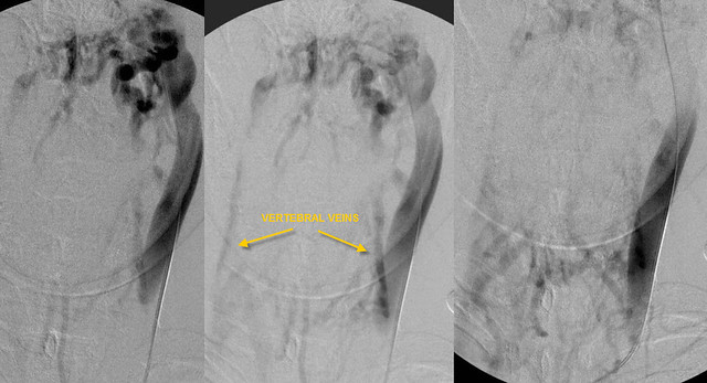
3. The emissary vein collateral... my anatomy book doesn't show it traversing the hypoglossal canal. The only pic I can find of the emissary vein is superior to the saggital sinus (Netter Atlas, plate 99 and 101).
Look for images of the cranial canals. anything that can transmit a nerve can have a vein travel along,

the following emissary veins are mentioned

mastoid emissary vein
thru mastoid foramen: transverse sinus to posterior auricular or occipital vein
parietal emissary v
thru parietal foramen: superior sagittal sinus to scalp veins

rete canalis hypoglossi
thru hypoglossal canal: transverse sinus to vertebral v and deep cervical veins

condyloid emissary vein
thru condyloid canal transverse sinus to deep veins of the neck

rete foraminis ovalis
thru foramen ovale cavernous sinus to pterygoid plexus

emissary vein of foramen of Vesalius and foramen lacerum
cavernous sinus to pterygoid plexus

internal carotid plexus
thru the carotid canal: cavernous sinus to IJV

Superior sagittal sinus with the veins of the nasal cavity.
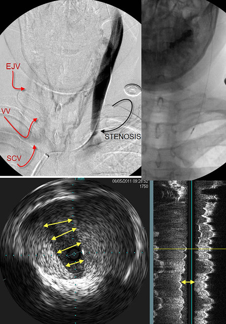
4. How could the contrast dye possibly illuminate the right IJV? A previous commenter hypothesised that the right IJV does not exist because it was not illuminated by the refluxed dye injected into the left IJV. Do the right and left IJV's communicate throught the emissary veins?
the emissary veins connect the dural sinuses to the extracranial veins by emissary veins that travel through the bony skull
5. I was about to hit the "submit" button, but saw the "mastoid emissary" in the general vicinity of the jugular bulb (the condylar emissary vein is also nearby). If either of these are the referenced collateral, please ignore #3 (but which one?). If not, please explain my confusion.
please share my confusion
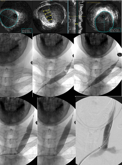
6. How much radiation is the patient exposed to during a nominal procedure?
7. How much are you and your staff exposed to radiation during a nominal procedure?
i am getting actual numbers for all my cases. this will take a while.
These are random, simple (blurted) questions. I am fascinated by this case, and I hope to have more and better questions soon. I look very much forward to hearing more about this.

Mike
I am sure you do