This Is MS Multiple Sclerosis Knowledge & Support Community
Welcome to This is MS, the leading forum for Multiple Sclerosis research and support. Join our friendly community of patients, caregivers, and researchers celebrating over 20 years of delivering hope through knowledge. https://www.thisisms.com/forum/
drsclafani wrote:IVUS, not shown here, revealed that there were internal echoes that represented scartissue or residual remnants of the valve or residual fixed valvular tissue.
Can you think of any way to differentiate between scar tissue or residual valve material? Would you expect any difference in immediate or long-term results when scar tissue is ballooned, compared to when residual valve tissue is ballooned?
Would a program of neck stretches potentially relieve some muscular compression? My son had infantile torticollis, which involves the same muscles that seem to be involved in CCSVI muscular compression cases. We did ear-to-the-shoulder stretches on him and then rotation stretches as if he was looking over his shoulder, twice a day.
Re: DrSclafani answers some questions
Posted: Wed Dec 14, 2011 9:33 am
by PaulH
drsclafani wrote:
Cece wrote:
drsclafani wrote:The "messy" area looks like reflux under an obstructed valve to me.
You had described this, using a wine glass in a tumbler image, at the symposium patient day. Here was me trying to describe what you had said: http://www.thisisms.com/forum/chronic-c ... ml#p171050
I bet there's a link out there for the actual presentation now....
so lets review the right internal jugular venograms. I will review all the IVUS studies together later:
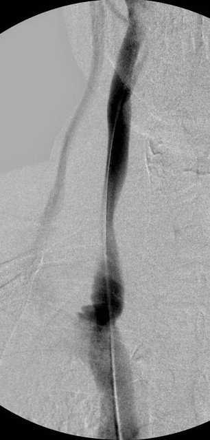
This vein was treated successfully a year ago. This image shows that there is a narrowing of the mid right IJV. Below this there is some irregularity and bulging of the contrast at the area ofof previously treated valve problems. There is reflux into the right external jugular vein (to the left of the IJV on the image).
Now a composite summary of the right side.
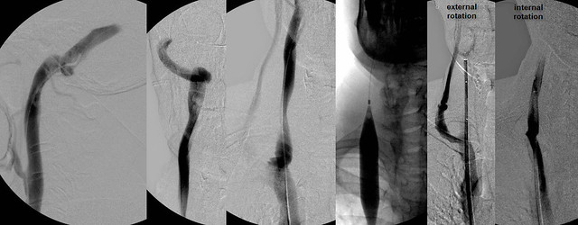
The first two images show the right transverse sinus emptying into the internal jugular vein. I think that this is the optimum way to fully evaluate the internal jugular vein. Opacify it by putting the constrast media upstream from the jugular vein. Sometimes, not often, you will see something wrong with the dural sinuses. You will also get a long at the condylar emissary vein which is a often seen collateral vein.Collaterals seen are minimal.
There was evidence of stenosis at the confluens where the valve was treated previously. There is refluxing contrast media in the external jugular vein and there is irregularity of the confluens. IVUS, not shown here, revealed that there were internal echoes that represented scartissue or residual remnants of the valve or residual fixed valvular tissue.
angioplasty was performed at the valvular area (balloon inflated). Then on to evaluate the mid jugular narrowing. Based upon this location, one should consider a few explanations.
1. Perhaps the first angioplasty was extended too high and the normal vein was overstretched and subsequently got scarred down. Not the case this time...
2. Consider that there was compression of the vein by the carotid artery.IVUS showed that this was not the case.
3. Final possibility was muscular compression. The final two images are with the neck rotated, first internally rotated, and the second one externally rotated. You can see that internal rotation results in a phasic complete compression of the vein with no flow at all in the IJV. All the contrast media was directed toward the external jugular vein. . When the head is turned externally, there is no stenosis and there was rapid flow.
Based upon this observation, one can say that this was a phasic narrowing caused by pressure of the muscles of the neck. I would advise no treatment beyond the angioplasty of the confluens' residual tissue narrowing the vein. Dilating muscle compression is unlikely to be helpful. Stenting is a possibility but I prefer NOT to place stents in the jugular except in a few circumstances.
Dr. Sclafani,
(not meaning to hijack the thread) But I wanted to let you know that I'm doing great since you inserted that 8cm stent into my right jugular vein two weeks ago. No fatigue. All is very well.
Thanks again for being such a pioneer in this field.
Paul.
Re: DrSclafani answers some questions
Posted: Wed Dec 14, 2011 9:36 am
by mo_en
Tempted to say the stent looks rather large compared to the phenomenal caliber of the RIJV. Seems like a candidate for the rendezvous procedure.
Re: DrSclafani answers some questions
Posted: Wed Dec 14, 2011 6:24 pm
by drsclafani
NZer1 wrote:So reading between the lines there is 'chance' that residual tissue will require attention, for some?
absolutely, poassible that residual valvular elements can fuse to themselves with tim. I really do not think that there will be very many patients who will have only one treatment. This will be an ongoing problem. My goal is to prolong treatment intervals.
Re: DrSclafani answers some questions
Posted: Wed Dec 14, 2011 6:28 pm
by drsclafani
pelopidas wrote:
drsclafani wrote:
3. Final possibility was muscular compression. The final two images are with the neck rotated, first internally rotated, and the second one externally rotated. You can see that internal rotation results in a phasic complete compression of the vein with no flow at all in the IJV. All the contrast media was directed toward the external jugular vein. . When the head is turned externally, there is no stenosis and there was rapid flow.
Based upon this observation, one can say that this was a phasic narrowing caused by pressure of the muscles of the neck. I would advise no treatment beyond the angioplasty of the confluens' residual tissue narrowing the vein. Dilating muscle compression is unlikely to be helpful. Stenting is a possibility but I prefer NOT to place stents in the jugular except in a few circumstances.
what are the treatment options?
thank you for the image uploading trick
and this is a very important lecture
pelopidas
thanks
why should we treat something that is not constant. afterall, venous drainage from the jugular veins is absent when the patient is erect.
Re: DrSclafani answers some questions
Posted: Wed Dec 14, 2011 6:33 pm
by Cece
drsclafani wrote:pelopidas
thanks
why should we treat something that is not constant. afterall, venous drainage from the jugular veins is absent when the patient is erect.
My understanding is that jugular flow is not absent when erect, but reduced by 90%?
Have you ever seen a patient where muscular compression is the only problem? No valve issues or hypoplasias or anything but muscular compression? I am guessing this is an unlikely scenario.
Re: DrSclafani answers some questions
Posted: Wed Dec 14, 2011 6:34 pm
by drsclafani
Cece wrote:
drsclafani wrote:IVUS, not shown here, revealed that there were internal echoes that represented scartissue or residual remnants of the valve or residual fixed valvular tissue.
Can you think of any way to differentiate between scar tissue or residual valve material? Would you expect any difference in immediate or long-term results when scar tissue is ballooned, compared to when residual valve tissue is ballooned?
i do not think it is going to matter at all. it is just important to find these problems when interventionis done. If venography cannot find them, the we need to find them so me other way. Perhaps it will be IVUS, perhaps optical tomography. but certainly we need to find it, otherwie problems will remain.
Would a program of neck stretches potentially relieve some muscular compression? My son had infantile torticollis, which involves the same muscles that seem to be involved in CCSVI muscular compression cases. We did ear-to-the-shoulder stretches on him and then rotation stretches as if he was looking over his shoulder, twice a day.
good question, cece.
first we need to understand the magnitude of the problem and then see if an intervention will reduce those problems. Then finally we need to assess further
Re: DrSclafani answers some questions
Posted: Wed Dec 14, 2011 6:45 pm
by drsclafani
PaulH wrote:
Dr. Sclafani,
(not meaning to hijack the thread) But I wanted to let you know that I'm doing great since you inserted that 8cm stent into my right jugular vein two weeks ago. No fatigue. All is very well.
Thanks again for being such a pioneer in this field.
Paul.
spectacular paul.
so interesting. i will share pauls story as he remains anonymous.
Delightful it is to hear about the improvement in symptoms
paul was treated with resolution of most major problems. then a few months later things reversed. Prompt ultrasound confirmed a problem with ccsvi and an occlusion was diagnosies. This was about four months after treatment but nonetheless, symptoms worsened quickly was an indication for aggressive followup
aggressive was I in addressing this deterioration of symptoms, within two weeks venography showed occlusion and i was able to do a rendevous procedure from above, open up the vein and stent an occlusion. If i had waited a few months (which i have in a few other cases), recanalization would have been doomed to failiure
the case very nicely illustrates the need for aggressive management of return of symptoms by ultrasound and venography.
Had i waited for a six month check, the vein would likely be lost and recanalization would have been impossible.
Donot wait passively by.
I am pleased that my patients provide me with good periodic followup. Those who are unable to travel to meet me on a routine basis, are asked to provide me with an email monthly. I pick up quite a few things this way.
I think i am using old and new ideas.
Re: DrSclafani answers some questions
Posted: Wed Dec 14, 2011 11:39 pm
by pelopidas
drsclafani wrote:[
very interesting hypothesis regarding reduction of neck pain.what about this consideration: patients with MS often havfe very large condylar emissary veins that connect to the veins in the posterior neck. Could relieving the resistance in the IJV, reduce flow through the emissary vein and thus reduce venous congestion in the neck muscles?
Sal
i guess this must be true, Dr Sclafani.
Very well and accurately described.
Let's name it the Sclafani Syndrome.
Re: DrSclafani answers some questions
Posted: Thu Dec 15, 2011 5:45 am
by drsclafani
mo_en wrote:Tempted to say the stent looks rather large compared to the phenomenal caliber of the RIJV. Seems like a candidate for the rendezvous procedure.
ok, now the shocker!
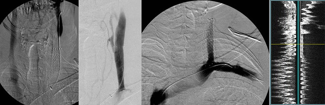
The left IJV is patent and has flow. The problem is that the flow is going from the left arm into the jugular vein and then from there to the intracranial dural sinuses and finally my patient's veins.
The image on the left has already been viewed. The second image with contrast injected within the stent showed that there was a smooth contour of contrast throughout the stent. But minimal contrast below the stent. The contrast column in the jugular vein is adequate, although the space between the edge of the stent and the contrast is in-stent intimal hyperplasia. The third image was an injection with the catheter within the subclavian vein. That vein which collects the blood from the left arm and the left chest wall has contrast that extends UP the left IJV, across the head and down the right IJV. So there just was NO downward flow in the IJV.
What is the explanation?
Re: DrSclafani answers some questions
Posted: Thu Dec 15, 2011 6:20 am
by mo_en
Its seems there is an obstacle inside the subclavian vein, forcing the blood upwards.
Re: DrSclafani answers some questions
Posted: Thu Dec 15, 2011 10:27 am
by Cece
Ok, that is a surprise! There is flow in the jugular but it is upward flow. Wrong direction. Such are the tricks our CCSVI veins play....
It's reminiscent of the subclavian stenosis that we saw previously, which was due to chemotherapy damage. This would appear to be an innominate vein stenosis. With no chemotherapy in the patient's history, perhaps it is due to a thrombosis. The guidewire and catheter were able to access the vein, so it is not a full occlusion. Treatment options would be ballooning followed by a stent?
I am presuming that during the prior procedure this stenosis was missed. The other possibility would be that the innominate vein stenosed after that procedure.
Re: DrSclafani answers some questions
Posted: Thu Dec 15, 2011 10:59 am
by Cece
drsclafani wrote:absolutely, poassible that residual valvular elements can fuse to themselves with tim. I really do not think that there will be very many patients who will have only one treatment. This will be an ongoing problem. My goal is to prolong treatment intervals.
You have a track record of being right....
I have heard from another doctor who posts in social media that 'done in one' is the goal. This is in contrast with your opinion here.
'Done in one' is nice if it happens, but realistically it has not been the experience of many thus far, which may not be a treatment failure but merely the nature of the beast we are up against....
drsclafani wrote:aggressive was I in addressing this deterioration of symptoms, within two weeks venography showed occlusion and i was able to do a rendevous procedure from above, open up the vein and stent an occlusion. If i had waited a few months (which i have in a few other cases), recanalization would have been doomed to failiure
Congrats to Paul, and to you, for this success.
Re: DrSclafani answers some questions
Posted: Thu Dec 15, 2011 1:15 pm
by pelopidas
drsclafani wrote: So there just was NO downward flow in the IJV.
What is the explanation?
is this stenosis permanent? could muscles restrict the vein ?
Re: DrSclafani answers some questions
Posted: Thu Dec 15, 2011 7:27 pm
by drsclafani
NZer1 wrote:So reading between the lines there is 'chance' that residual tissue will require attention, for some?
its the nature of veins that retreatment is the rule
it is no different than getting a flu shot doesnt last forever, no different than getting a tooth cavity fixed and getting more tooth decay. its like so many other disease. we manage, we do not cure