Page 452 of 557

Re: DrSclafani answers some questions

Posted: Sat Apr 28, 2012 4:52 pm
by drsclafani
Donnchadh wrote:This is something I have been wondering about: is it possible to utilize collateral veins as a substitute IJV? There have been some cases of people with congenital deformed, or severely undersized, IJV's which might not respond to having them dilated. Also, some patients have severely scarred IJV which cannot be treated. I realized that each case is different, but let's suppose there are a network of collateral veins running roughly parallel to the IJV.

Would it be possible to enlarge a target collateral vein (and maybe re-routing them to facilitate flow) to function as a IJV?

Donnchadh
yes, donnchadh
it would be possible. I have already dilated stenotic valves of collaterals to improve flow.There are collaterals such as the facial vein and anterior and posterior jugular veins which enlarge whenever there is occlusion or stenosis of the IJV. However these veins are not as large as the internal jugular veins and just dont do the job of the IJV itself.

Re: DrSclafani answers some questions

Posted: Sat Apr 28, 2012 4:58 pm
by drsclafani
Donnchadh wrote:This is something I have been wondering about: is it possible to utilize collateral veins as a substitute IJV? There have been some cases of people with congenital deformed, or severely undersized, IJV's which might not respond to having them dilated. Also, some patients have severely scarred IJV which cannot be treated. I realized that each case is different, but let's suppose there are a network of collateral veins running roughly parallel to the IJV.

Would it be possible to enlarge a target collateral vein (and maybe re-routing them to facilitate flow) to function as a IJV?

Donnchadh
When there is occlusion or stenosis of the IJVs, it is common for collateral veins to take over the flow. The facial vein, the condylar emissary veins, the anterior and external jugular veins are common enlarged when there is long term outflow of obstructions of the IJVs.

I have performed valvuloplasty of such veins when hypoplasia, or occlusion have occured. However these veins are smaller than the Normal IJV and they are tortuous. thus flow is never as substantial as the good old IJV

Re: DrSclafani answers some questions

Posted: Sat Apr 28, 2012 5:12 pm
by drsclafani
drsclafani wrote:After several hundred procedures, truly unusual and enigmatic procedures sometimes come along. Despite difficulties in diagnosis, an unusual angiographic appearance of uncertain cause, and a lack of clarity of the pathology, a great outcome comes from limited interventions.
The patient is a 42 year old Canadian who was diagnosed with secondary progressive multiple sclerosis. He had had symptoms of neurological disease for more than twenty years beginning with "flashing lights in his head and grand mal seizures as a young adult. For years no diagnosis was made. The patient was told that it was "nerves", or anxiety. In June 2011 he was finally diagnosed with progressive multiple sclerosis.
Currently he walks with a cane except for long distances which require a wheelchair, having weakness in his left leg, a foot drop and imbalance that it is worst at night. He suffers from heath intolerance and intense fatigue: he sometimes sleeps for two days. He has numbness and tingling in both torso, hands and feet. He has headaches and electric shocks in his left leg.
On examination, he had difficulties with memory, decision making and word and number recall. Cranial nerves exam revealed positive Rhomberg test and weakness of spinal accessory muscles. He had weakness in left left hip function, and paralysis of his left ankle and foot. He had absent sensation to vibration and light and sharp touch in his left fingers and toes and the dorsum of his left foot. He could not walk without a cane.
His Doppler examination had three criteria of CCSVI. The valve leaflets of the right and left internal jugular veins were extremely long. Reflux was noted in both jugulars and the deep cerebral veins.
I performed venography last week.
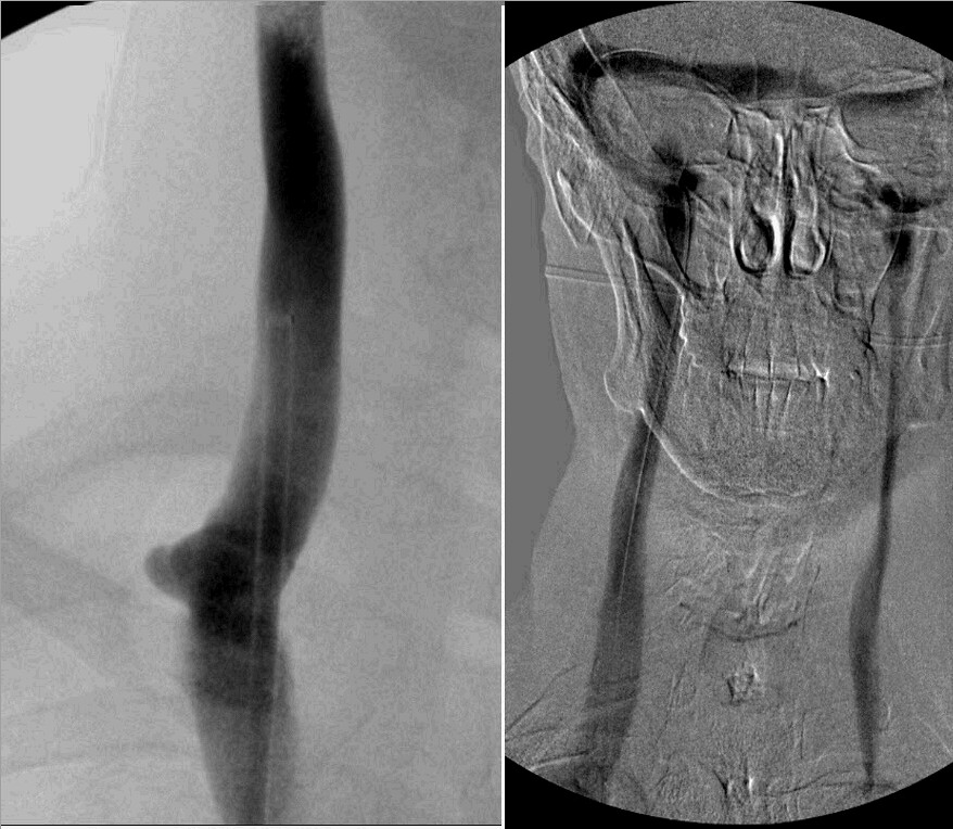
Let's start with the right IJV

Image

Re: DrSclafani answers some questions

Posted: Sat Apr 28, 2012 6:47 pm
by tiltawhirl
drsclafani wrote:
Cece wrote:Any interesting cases lately?
yes, i started posting a new case above
;)

tilt

Re: DrSclafani answers some questions

Posted: Sat Apr 28, 2012 8:50 pm
by Cece
drsclafani wrote:Let's start with the right IJV

Image
It looks like there is that bump in the left image, with some dark contrast at that area, but it doesn't appear to be narrowed. In the right image, the right jugular looks wide and good at the area of the valves. But where is the contrast being injected in that right image? From the image it looks like the catheter is snaked through the sinuses into the left jugular, and that there is no flow within the left jugular, and the contrast is going down a collateral that could be mistaken for a jugular as well as back through the sinuses to the right jugular. Is that accurate, or am I seeing things?

Re: DrSclafani answers some questions

Posted: Sun Apr 29, 2012 12:30 am
by NZer1
Dr.S,
I have been approached by a TV producer to answer some questions;
Chris Cooke
Hi I am a prodcuer with SUNDAY at TVNZ. You will have seen our story from overseas on the liberation treatment. I would like to make contact with New Zealnders looking to travel overeas for the proccedure in the near future. Can you assist?
The clip from Australia;
http://www.facebook.com/l.php?u=http%3A ... s__Uglo03A

I have been a little quiet of late, MS thing, but I seem to have fired up.
At the moment there is news down under of the Alfred Hospital (Australia) getting their FDA approval for a study. I have been in contact a bit with Dr. Paul Thibault about my CPN positive and in the discussions we talked about IVUS use by IR's in Australia. Bottom line is that they are not that well trained on the benefits and not that aware of the learning curve available in CCSVI if they use IVUS.

*So do you have any contact with the likes of the Alfred?

*What would your advice be that I could pass on to them regarding a study on CCSVI without the use of IVUS?

* The TV Producer is a work in progress as he hasn't called as he agreed to after he asked to speak in the first instance. I am going to make contact during work hours as he may be one of those who keep to those constraints. Is there anything that in your opinion needs to be aired?

*It has fired me up to travel and say hello, mostly because I am going down hill a little faster lately, so I am wanting to help the cause if I can with the TV producers interest being active. Sadly my sister has just left for the States, but, I do have a friend who is willing to travel with me as he has a son training in Aeronautics over your way at the moment and is travelling back and forth allot and he is also a producer/photographer in Australia.

Regards Nigel

Re: DrSclafani answers some questions

Posted: Sun Apr 29, 2012 12:50 am
by gibbledygook
Dear Professor Sclafani,

Thank you for your tireless work in CCSVI and on this board. I am booked in to see you in December and am very interested in using helminth therapy (it seems pretty safe with weak adverse effects) but wondered if I could commence helminth therapy prior to having venoplasty possibly with stents. Do you think it would be safe to start hookworm therapy prior to surgery?
best wishes, Alex Gibbs

Re: DrSclafani answers some questions

Posted: Sun Apr 29, 2012 5:53 am
by drsclafani
gibbledygook wrote:Dear Professor Sclafani,

Thank you for your tireless work in CCSVI and on this board. I am booked in to see you in December and am very interested in using helminth therapy (it seems pretty safe with weak adverse effects) but wondered if I could commence helminth therapy prior to having venoplasty possibly with stents. Do you think it would be safe to start hookworm therapy prior to surgery?
best wishes, Alex Gibbs
Alex, i didnt find much data on helminth therapy except to that it is thought to reduce autoimmunity. I searched complications and did not find much either.

So i dcannot say that i can provide you with any advice regarding helminth therapy. You already have stents and i just cannot advise you in the positive regarding infecting yourself with parasites in light of the fact that you have metal in your veins already. How hookworms would interact with these devices is totally unreported. You should speak to a parasitologist about this.

Without definitive answers and with unclear benefits, i would not support you trying this before a vascular therapy.

DrS

Re: DrSclafani answers some questions

Posted: Sun Apr 29, 2012 5:59 am
by drsclafani
Cece wrote:
drsclafani wrote:Let's start with the right IJV

Image
It looks like there is that bump in the left image, with some dark contrast at that area, but it doesn't appear to be narrowed. In the right image, the right jugular looks wide and good at the area of the valves. But where is the contrast being injected in that right image? From the image it looks like the catheter is snaked through the sinuses into the left jugular, and that there is no flow within the left jugular, and the contrast is going down a collateral that could be mistaken for a jugular as well as back through the sinuses to the right jugular. Is that accurate, or am I seeing things?
Let me say that in the right image, the catheter is in the right transverse sinus. The vein on the left side that you call a collateral is in direct continuity with the dural sinus. That indicates that it is the internal jugular vein.

You can never say from a single image that there is no flow. it is like looking at a still frame from a movie, or for those too young to remember super 8 or 35 mm movies, a dvd on pause and saying it is not a movie.

Does anyone see a stenosis in the venogram on the left?

Re: DrSclafani answers some questions

Posted: Sun Apr 29, 2012 8:23 am
by drsclafani
NZer1 wrote:Dr.S,
ht
I have been a little quiet of late, MS thing, but I seem to have fired up.
At the moment there is news down under of the Alfred Hospital (Australia) getting their FDA approval for a study. I have been in contact a bit with Dr. Paul Thibault about my CPN positive and in the discussions we talked about IVUS use by IR's in Australia. Bottom line is that they are not that well trained on the benefits and not that aware of the learning curve available in CCSVI if they use IVUS.

*So do you have any contact with the likes of the Alfred?
I have had correspondence with Dr Thompson and invited him to my symposium in 2010 but he has not contacted me.
l

*What would your advice be that I could pass on to them regarding a study on CCSVI without the use of IVUS?
it is my opinion IVUS is necessary to identify intraluminal pathology such as webs, and immobile valves. Without it randomized studies will be flawed.

*It has fired me up to travel and say hello, mostly because I am going down hill a little faster lately, so I am wanting to help the cause if I can with the TV producers interest being active. Sadly my sister has just left for the States, but, I do have a friend who is willing to travel with me as he has a son training in Aeronautics over your way at the moment and is travelling back and forth allot and he is also a producer/photographer in Australia.

Regards Nigel
I look forward to meeting you nigel. you are always welcome.

Re: DrSclafani answers some questions

Posted: Sun Apr 29, 2012 2:00 pm
by Cece
drsclafani wrote:Does anyone see a stenosis in the venogram on the left?
Image
Some areas of interest:
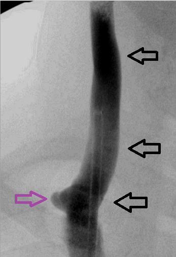
top black arrow points to the dark contrast
middle black arrow points to lighter contrast
bottom black arrow points to darkening of contrast
cute purple arrow points to a protrusion

I would guess that area between the purple arrow and the lowest black arrow is the stenosis because of the protrusion and the darker contrast. Even though it doesn't look narrowed. If the protrusion is contrast refluxing up under the flaps of a valve, why does it stick so far out from the vein itself? Dilatation of the vein at that area?

Or could the protrusion be reflux into the subclavian vein, and the stenosis is higher, between the top and middle arrows because of the contrast going from dark to light?

This isn't easy...

Re: DrSclafani answers some questions

Posted: Sun Apr 29, 2012 2:55 pm
by drsclafani
Cece wrote:
drsclafani wrote:Does anyone see a stenosis in the venogram on the left?
Image
Some areas of interest:
top black arrow points to the dark contrast
middle black arrow points to lighter contrast
bottom black arrow points to darkening of contrast
cute purple arrow points to a protrusion

I would guess that area between the purple arrow and the lowest black arrow is the stenosis because of the protrusion and the darker contrast. Even though it doesn't look narrowed. If the protrusion is contrast refluxing up under the flaps of a valve, why does it stick so far out from the vein itself? Dilatation of the vein at that area?

Or could the protrusion be reflux into the subclavian vein, and the stenosis is higher, between the top and middle arrows because of the contrast going from dark to light?

This isn't easy...
But you are brave to take a stab at it.

There is no evidence of stenosis. There is indirect evidence of obstruction.
1. there is reflux into the opposite internal jugular vein, despite the left IJV being smaller than the right IJV
2.The protrusion, as you call it, represents ballooning out of the undersurface of the valve area because of resistance at the obstruction.

So there is a stenosis but the exact location of that stenosis is not visible.

Where to angioplasty?

Re: DrSclafani answers some questions

Posted: Sun Apr 29, 2012 3:22 pm
by NZer1
IVUS, why guess?
Seems to me that visually we are seeing areas of interest only, follow the clues/hints and IVUS to remove doubt!

Re: DrSclafani answers some questions

Posted: Sun Apr 29, 2012 4:24 pm
by Cece
drsclafani wrote:2.The protrusion, as you call it, represents ballooning out of the undersurface of the valve area because of resistance at the obstruction.
Is it possible for the undersurface of the valve area to balloon outward like that if the obstruction is not the valve itself?
So there is a stenosis but the exact location of that stenosis is not visible.

Where to angioplasty?
I agree with Nigel, it is time to bring in the IVUS. But if this is to be done with angiography only, it would be a good guess to balloon the area of the valves, because most of the time, CCSVI is a malformation of the valves. Another possibility would be a septum located above the valve, if such a septum would suffice to cause that protrusion of the vein beneath the valve?

A septum would be an obstruction without being a stenosis.

Re: DrSclafani answers some questions

Posted: Sun Apr 29, 2012 8:22 pm
by drsclafani
NZer1 wrote:IVUS, why guess?
Seems to me that visually we are seeing areas of interest only, follow the clues/hints and IVUS to remove doubt!

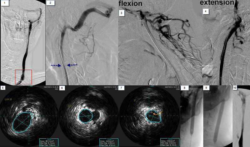
As you wish. my goal was to illustrate that stenoses are present even in the absence of direct evidence of stenosis by venography. Image
i think the image speak for themselves. IVUS shows immobile valves treated by 18 mm angioplasty to 8 ATM.

The left jugular venogram , as you could see from the right sided venogram reflux, is small in the upper area of the neck. Further it is narrowed even more in the lower neck. This valve was quite tight.

The initial work was done in the lower jugular vein. I wanted to assure myself that the upper narrowing wasn't just a physiological narrowing caused by the lower stenosis.
Image
There is a stenosis at the confluens with the subclavian vein (red arrows). there is reflux into the anterior jugular vein. images 3,4,5,6 show the thickened leaflets (white arrows) resulting in a 67% stenosis of this hypoplastic vein. These thicked areas are better seen in image 7. A 14 mm balloon was inflated to open this valve. The post angioplasty imag shows improvement in luminal diameter and no visualization of the anterior jugular vein.

Image

The upper jugular vein was more challenging. in image 2 you can see the narrowing of the upper internal jugular vein (blue arrows). By flexing the neck, the vein is completely occluded with a lot of filling of posterior cervical veins connecting to emissary veins. In extension, the lumen looks great!
The three ivus images show the dimensions of the vein. a 7mm vein decreased to a 4mm balloon lower down and it remained that size for a while. So, is this a hypoplasia? in image 7 the orange arrown points to intraluminal material. That resembles thrombus to me. Perhaps this is all a recanalized old occlusion of the IJV? or the results of an inflammatory process? The patient said he had many throat infections while growing up. Could this be the result of an infectious process? Any way, i did angioplasty with a long 8 mm balloon. REsults seem to show some improvements

Finally, the azygous vein, the brachiocephalic veins, the left renal vein, the left ascending lumbar vein, the left common and exernal iliac veins were all normal .

On the table the patient said that his pins and needles were gone and he started to move his paralyzed ankle. Said vision might be a bit clearer too.

In recovery room i reevaluated him. Strength in his paralyzed ankle was normal and equal to the other side. numbness in both hands and feet had resolved. unlike the day before, he could feel the pain of a n eedle in his foot and fingers. Rhomberg;s test was normal, he could stand with his eyes closed.,

He came out of the dressing room carrying his cane, not using it. Amazing result.

The next day he went on a two block walk without his cane.