Page 5 of 6
Posted: Thu Jul 01, 2010 3:22 pm
by L
Lyon wrote:L wrote:Maybe the answer is to shut down all of the sub forums except this one?
Good idea!
We could just call it "thisisccsvi" because that's pretty much what it is in everything but name.
That really is a great idea. Champion, as my granddad would have said.
Posted: Thu Jul 01, 2010 3:24 pm
by jimmylegs
semantics. this thread's 'debate' has had nothing to do with CCSVI merits or lack thereof.
yes it got reported. all TIMS members, regardless of 'side', are free to report rules violations when they occur.
to all readers, if you have thoughts on the presentation link that started this thread, i invite you to share your thoughts and comments, in line with the rules of the forum.
jimmy out
Posted: Thu Jul 01, 2010 3:50 pm
by Cece
jimmylegs wrote:to all readers, if you have thoughts on the presentation link that started this thread, i invite you to share your thoughts and comments, in line with the rules of the forum.
In the picture on the first page, Dr Sclafani is pointing to a screen that says 'Distension of the carotid impression'...anyone know what that means?
Posted: Thu Jul 01, 2010 4:16 pm
by cheerleader
Cece wrote:jimmylegs wrote:to all readers, if you have thoughts on the presentation link that started this thread, i invite you to share your thoughts and comments, in line with the rules of the forum.
In the picture on the first page, Dr Sclafani is pointing to a screen that says 'Distension of the carotid impression'...anyone know what that means?
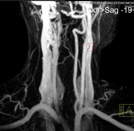
Yes, Cece. One of the ways in which the jugular can become compressed is by the carotid artery putting pressure on it. Like May-Thurner where the iliac vein is compressed by the iliac artery. Since veins are less rigid than arteries, they give way to the artery. Dr. Haacke reported on this finding first, since he saw it on MRV- you can see a 3D image under malformation of the internal carotid artery that compresses the internal jugular vein. This malformation could also be called a distension of the carotid.
http://www.ms-mri.com/video.php?filesent=artery%201.flv&
Re: Dr. Sclafani's Italy Presentation
Posted: Thu Jul 01, 2010 9:48 pm
by NHE
This thread is not the place to discuss the moderation of the forums. All further posts on this topic in this thread will be moved to a thread in the support forum.
Thank you for your cooperation, NHE
Posted: Thu Jul 01, 2010 9:56 pm
by Cece
Thanks Cheerleader. I'd read about that one awhile back but forgotten along the way. With MT being similar, there might be other veins in the body that can be affected by arteries in this way?
Posted: Sat Jul 03, 2010 3:47 am
by CureOrBust
cheerleader wrote:One of the ways in which the jugular can become compressed is by the carotid artery putting pressure on it. Like May-Thurner where the iliac vein is compressed by the iliac artery. Since veins are less rigid than arteries, they give way to the artery.
But what can be done about this? I can't see them placing a stent at the point the jugular crosses the carotid artery, as it may then cause a stenosis of the artery supplying the brain, and if ballooned, the artery will simply press against it again. Or am I well off the mark here?
And just to be extra clear, is this the sort of thing they are talking about?

Posted: Sat Jul 03, 2010 6:43 am
by Algis
If it can be fixed in the May-Thurner syndrome; a vein is a vein and artery is an artery (?)
But do not take my word for it

Posted: Sat Jul 03, 2010 7:29 am
by Cece
My guess is that a stent would be the solution. The blood flows through the carotid artery at high pressure, I do not think the 'stent next door' would be a problem for it, unless there is something entrapping the carotid on the other side.
Posted: Tue Jul 06, 2010 9:35 pm
by drsclafani
cheerleader wrote:Cece wrote:jimmylegs wrote:to all readers, if you have thoughts on the presentation link that started this thread, i invite you to share your thoughts and comments, in line with the rules of the forum.
In the picture on the first page, Dr Sclafani is pointing to a screen that says 'Distension of the carotid impression'...anyone know what that means?
Yes, Cece. One of the ways in which the jugular can become compressed is by the carotid artery putting pressure on it. Like May-Thurner where the iliac vein is compressed by the iliac artery. Since veins are less rigid than arteries, they give way to the artery. Dr. Haacke reported on this finding first, since he saw it on MRV- you can see a 3D image under malformation of the internal carotid artery that compresses the internal jugular vein. This malformation could also be called a distension of the carotid.
http://www.ms-mri.com/video.php?filesent=artery%201.flv&
Actually everyone has misinterpreted the meaning of the title of the slide.
The slide is showing the value of IVUS. The Carotid IMPRESSION on the jugular vein is caused by carotid compression of a jugular vein that has outflow obstruction more distally near the confluens with the subclavian vein. I am trying to prove by using IVUS in each case whether this carotid impression is transient and physiological, or pathological.
The goal of the images in this example was to show that IVUS could prove that this was not a fixed stenosis. The image on the left and right are of the same area of the same vein in two states: NARROWING by the carotid impression on the left at rest.....and DISTENSION of the carotid impression on the right during activation of the thoracic pump in the image. In this case, the information from IVUS was used to avoid "correcting" a physiological narrowing without the need for angioplasty or stenting.
Posted: Tue Jul 06, 2010 9:41 pm
by drsclafani
Cece wrote:My guess is that a stent would be the solution. The blood flows through the carotid artery at high pressure, I do not think the 'stent next door' would be a problem for it, unless there is something entrapping the carotid on the other side.
I call things like the carotid impression...."leave me alone lesions".
stent is not what i want to put there.
my concerns are severalfold
1. stenting a physiological narrowing may be a problem when that narrowing distends by treatment of lower more likely stenoses. it is possible that the stent will no longer be pressed against the wall of the vein and thus migrate toward the heart.
2. stents have the risks of in-stent stenosis leading to loss of the vein completely...certainly do not want to put one there for a physiological distensible narrowing
3. putting pressure on the wall of the carotid at the carotid bulb by stenting the vein may result in physiological responses including a slow heart rate
Posted: Tue Jul 06, 2010 9:46 pm
by Cece
This is why you are the IR and not me! I retract my decision to stent and am glad I am not in charge of making any such actual decisions.
drsclafani wrote:The slide is showing the value of IVUS. The Carotid IMPRESSION on the jugular vein is caused by carotid compression of a jugular vein that has outflow obstruction more distally near the confluens with the subclavian vein. I am trying to prove by using IVUS in each case whether this carotid impression is transient and physiological, or pathological.
The goal of the images in this example was to show that IVUS could prove that this was not a fixed stenosis. The image on the left and right are of the same area of the same vein in two states: NARROWING by the carotid impression on the left at rest.....and DISTENSION of the carotid impression on the right during activation of the thoracic pump in the image. In this case, the information from IVUS was used to avoid "correcting" a physiological narrowing without the need for angioplasty or stenting.
I will reread this in the morning, but I think I've got it. Fascinating stuff.
Posted: Wed Jul 07, 2010 4:22 am
by Nunzio
I like to make an important point in understanding CCSVI.
An artery or bone cannot indent the Jugular vein.
The only reason that we think so is that is what we see in the pictures.
In reality veins are very soft and pliable and can easily go around fixed obstructions.
Veins can only be squeezed between two structures.
So when we see a vein being indented we have to think about what is pushing the vein against the artery or bone.
The implication is that ballooning such a vein will not achieve anything because the vein will still be squeezed between the two structures.
According to Dr. Noda, the neck muscles are the missing link in this story.
So, in the future please say " the jugular vein is squeezed between the carotid artery and ........"
This will further our understanding of what is really impairing our blood flow.
Posted: Wed Jul 07, 2010 8:06 am
by Cece
In this particular situation I do not think the vein was squeezed between two structures, since it was able to expand under valsalva as seen using ivus.
In May Thurner, when the iliac artery is lying on the iliac vein and compressing it, what is on the other side? Is it being squeezed? Why is MT not a leave-me-alone lesion...I suppose because it is nowhere near the heart and brain!
VI
Posted: Wed Jul 07, 2010 11:56 am
by Nunzio
Cece wrote:In this particular situation I do not think the vein was squeezed between two structures, since it was able to expand under valsalva as seen using ivus.
In May Thurner, when the iliac artery is lying on the iliac vein and compressing it, what is on the other side? Is it being squeezed? Why is MT not a leave-me-alone lesion...I suppose because it is nowhere near the heart and brain!
It is
always squeezed between two structures.
In May Thurner syndrome the Iliac vein is between the Iliac artery and the backbone. Click on the link and you will see the anatomy.
http://radiology.rsna.org/content/233/2 ... nsion.html
For the Jugular vein I think is a muscle gently squeezing the vein. Valsalva can overcome the compression but this only means it is not an intrinsic narrowing but a narrowing from the outside compression.
Since CCSVI is a problem with flow I think in the above case a stent would help overcame the mild compression causing the still significant narrowing.
This is what they do in Poland where they do not have an IVUS yet anyway.
Only more testing will clarify this point and maybe lead to alternative treatment.
If you look at your arm and hand, you can see that the veins do not have any problems going around the bones and tendons present there.
