Page 6 of 7
Posted: Thu Dec 09, 2010 9:32 pm
by Cece
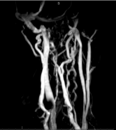
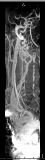
It's pretty obvious now, on both sides, where the jugulars come to a near-complete stop.
I've looked for comparison images online and the one from the ISNVD (international society of neurovascular diseases) seems closest:
http://www.isnvd.org/
Posted: Fri Dec 10, 2010 8:53 am
by Cece
Now that I figured out how to add images

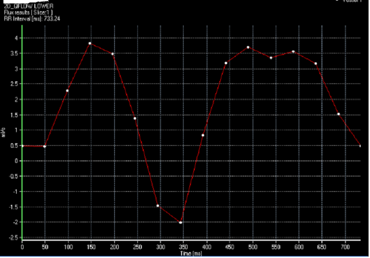
here's what I've been talking about with the flow graph:
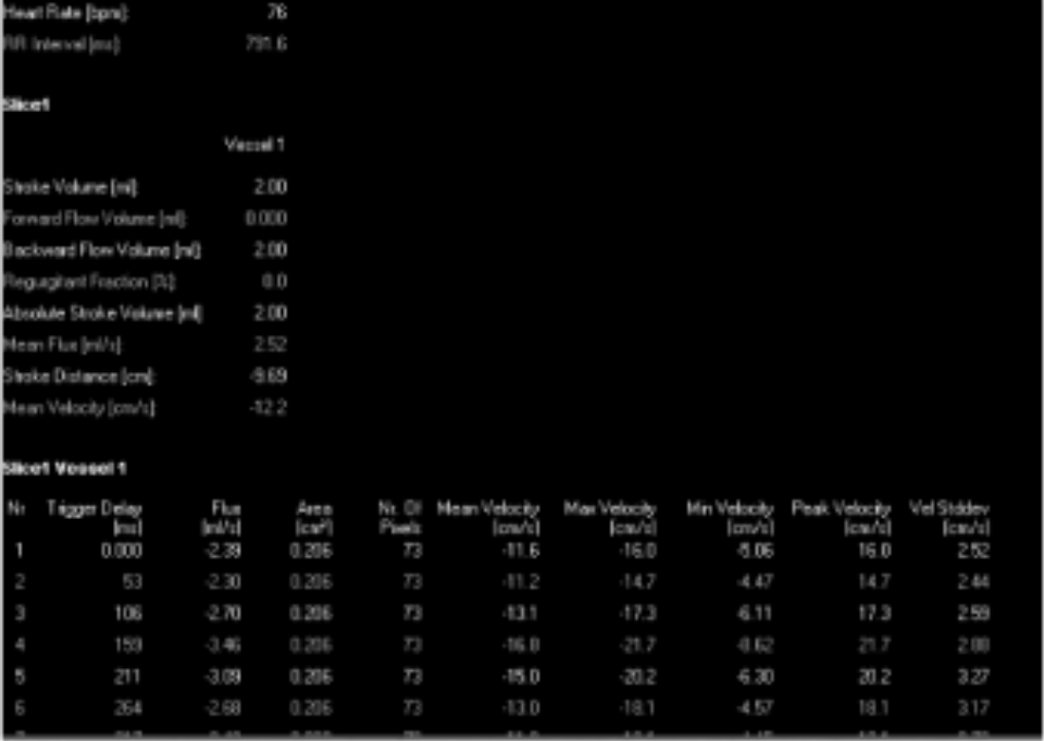
Forward flow volume: 0
Backward flow volume (ml): 2.00
Regurgitant flow: 0 *
Stroke distance (cm): -9.69
Mean velocity (cm/s): -12.2
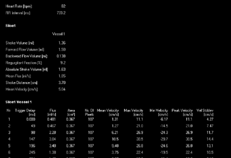
* in my azygous, I have a number here for regurgitant flow, because in the azygous there is both forward and backward flow
This is my right jugular but the numbers for my left are nearly identical.
Posted: Fri Dec 10, 2010 9:00 am
by Cece
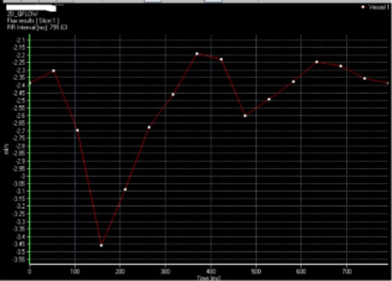
The graph too.
These are a little big, sorry about that, still learning.

Posted: Fri Dec 10, 2010 12:52 pm
by Sotiris
According to the following report
http://www.falsecreekhealthcare.com/sha ... n-2010.pdf your jugulars seem to have no reflux (see page 11 where it is stated that "The right IJV has slightly positive flow at the end of the cardiac cycle, indicating minor reflux.", i.e. backward flow means flow back to heart). I think that this is also the meaning of "Regurgitant fraction = 0%". Reflux seems to be a problem with your azygos vein.
Good luck again with your procedure in January
Posted: Fri Dec 10, 2010 1:37 pm
by Cece
Sotiris wrote:According to the following report
http://www.falsecreekhealthcare.com/sha ... n-2010.pdf your jugulars seem to have no reflux (see page 11 where it is stated that "The right IJV has slightly positive flow at the end of the cardiac cycle, indicating minor reflux.", i.e. backward flow means flow back to heart). I think that this is also the meaning of "Regurgitant fraction = 0%". Reflux seems to be a problem with your azygos vein.
Good luck again with your procedure in January
thanks for the link, Sotiris, I will read through it.
AZYGOUS
Posted: Fri Dec 10, 2010 1:58 pm
by Cece
azygous
Posted: Fri Dec 10, 2010 1:59 pm
by Cece
Posted: Fri Dec 10, 2010 2:14 pm
by Cece
Sotiris, do you know what this means? I am so confused about which direction positive and negative is. It's from one of the last pages in the document you linked to.
For Siemens data, the flow away from the heart to the brain (typically arterial flow) is defined as the positive direction, which shows up as white vessels in the phase image. The whiter it is, the higher the
velocity. The flow toward the heart (typically venous flow) is in the negative direction, which shows up as dark vessels in the phase image. The darker it is, the higher the velocity toward the heart. For GE flow quantification data, the positive and negative directions are reversed.
Is what I have Siemens data or GE flow quantification?
Posted: Fri Dec 10, 2010 2:31 pm
by Sotiris
Cece wrote:Sotiris, do you know what this means? I am so confused about which direction positive and negative is.
For Siemens data, the flow away from the heart to the brain (typically arterial flow) is defined as the positive direction, which shows up as white vessels in the phase image. The whiter it is, the higher the
velocity. The flow toward the heart (typically venous flow) is in the negative direction, which shows up as dark vessels in the phase image. The darker it is, the higher the velocity toward the heart. For GE flow quantification data, the positive and negative directions are reversed.
Is what I have Siemens data or GE flow quantification???
I'm not sure about which machine they have used in your case, but it is quite clear from the term "
regurgitant fraction %" that in your case the flow in the azygos vein has to be positive (9.2%=0.138/1.5). In the chart the backward flow 0.138ml volume corresponds to the area that is below the 0 line, while the 1.5 ml volume corresponds to the area above the 0 line.
Posted: Fri Dec 10, 2010 3:20 pm
by Cece
thanks, Sotiris, I'll read this through a few more times, much appreciated.
Posted: Fri Dec 10, 2010 3:21 pm
by Cece
Here's another angle on one of my jugulars:
Posted: Fri Dec 10, 2010 3:59 pm
by JohnJoseph
Cece,
missing the confluence with subclavian - do you have images of that crutial area with valves?
JJ
Posted: Fri Dec 10, 2010 4:34 pm
by Cece
JohnJoseph wrote:Cece,
missing the confluence with subclavian - do you have images of that crutial area with valves?
JJ
I think I do but I don't really know what I'm seeing so don't know if I'm catching it at a good angle (it rotates in 3D).
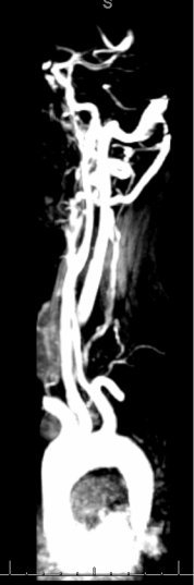
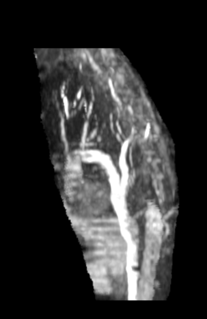
(left jugular)
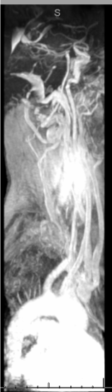
(left jugular again)
Do you see anything, John Joseph? It doesn't even look like a left jugular to me anywhere in there.
edited to add: ok,if my left jugular is the foreshortened thing in the first picture, it looks like in the second picture the contrast has passed through all other veins except for the very bright dead-end jugular what might be a neighboring collateral that is also very bright. The jugular itself is difficult to see beneath that point, maybe it does not exist?

Posted: Fri Dec 10, 2010 4:56 pm
by Cece
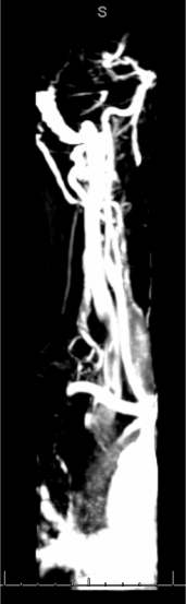
(right jugular)
(right jugular again)
Posted: Fri Dec 10, 2010 5:19 pm
by Cece
last one:
my azygous, rotated, to show the black indent down low on it?