Page 40 of 49
Posted: Wed Jul 20, 2011 2:55 pm
by HappyPoet
Cece wrote:Ok, no changes in my numbness in my left for 8 days post-procedure and then in a matter of hours it melts away entirely.
This is a numbness that comes and goes but it does not go in this fashion, except once before, after my first venoplasty. I am sufficiently awed.
So happy for you, Cece

I'll be hoping more improvements keep coming and this procedure lasts a nice, long time

Posted: Wed Jul 20, 2011 3:01 pm
by Cece
Thanks, HappyPoet. I am so glad we got to meet and talk at the symposium!! You have been a friend here at TIMS from the day I arrived.
Posted: Wed Jul 20, 2011 3:05 pm
by Cece
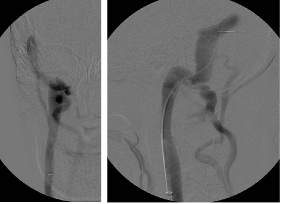
Here is my dural sinus venogram on the right side....
Posted: Wed Jul 20, 2011 3:25 pm
by Cece
Here is my left side....
Posted: Wed Jul 20, 2011 3:37 pm
by Cece
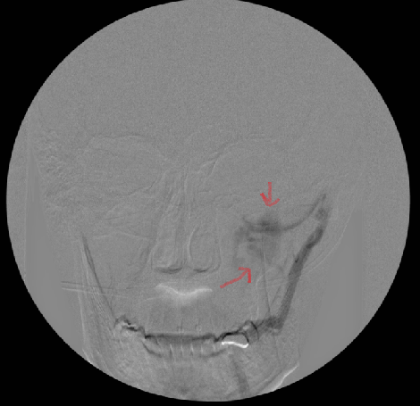
ok I drew some arrows on these....
I think the blue arrow is pointing to the dural sinus, where the contrast was released. The orange arrow is pointing to the jugular. The light green arrow is pointing to the vertebral veins? These should not be as darkly lit as they are. When he saw them light up like that, that was when he knew there was a blockage lower down, even though he was not at that time looking at the area of the valves. The red arrows are pointing to the squiggle of veins that also should not be lighting up like that.
Posted: Wed Jul 20, 2011 4:06 pm
by Cece
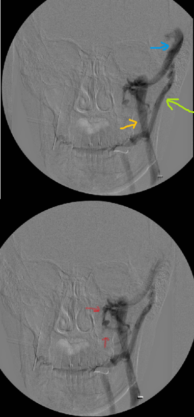
almost forgot this one...
This is my left dural sinus from the side angle. The sinus itself looks healthy, as did the sinus on my right side. It is connected to the jugular as it should be.
Any questions? ;)
Posted: Thu Jul 21, 2011 10:15 am
by Cece
Here is something else of interest. This is not of me. It comes from this site:
http://www.musc.edu/intrad/AtlasofVascu ... 6FIG1A.jpg
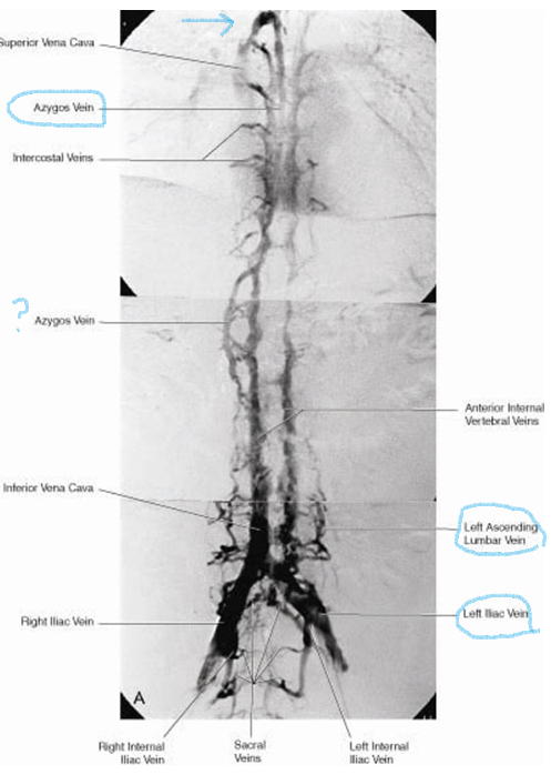
I found it helpful to see it all in one image like this. These are veins along the spine. At the top you see our good friend the azygos vein. I drew an arrow to the arch, which is where problems mostly are. There's another labelled azygos vein lower down, what is that about? Is it the hemiazygos? Or is still the azygos?
Lower down I circled the ascending lumbar veins, which are sometimes malformed in patients with MS but are too small to treat, but Dr. Zamboni still chose to image them. Also circled is the left iliac vein. When the left iliac vein is compressed, that is called May Thurner syndrome, which we have all heard plenty about around here.
Posted: Thu Jul 21, 2011 10:32 am
by Cece
from the same site:
http://www.musc.edu/intrad/AtlasofVascu ... P6FIG2.jpg

You can see the vulnerable spinal cord tissue. And all the veins surrounding it.
Posted: Thu Jul 21, 2011 10:40 am
by Cece
http://tinyurl.com/3swqsem
A dissection image. Left and right jugular veins and their connection with the subclavian vein are easily seen. This juncture with the subclavian is where the valves are located and, in CCSVI, these valves are often malformed and blocking flow.
I have to say, there is nothing on the internet about compression of the azygos by the aorta. That's what started me hunting. Although I have found other interesting things.
Posted: Thu Jul 21, 2011 3:45 pm
by HappyPoet
Cece, thanks for sharing and labeling your great images.
It looks like DrS brought the catheter into the Transverse Sinus (TS) just a bit before releasing the contrast dye. Very good news to learn your Sigmoid Sinuses (SSs) are properly connected to their respective IJVs with no SS stenoses. Very interesting to actually see an image of the vertebral veins (VVs) with dye going down them.
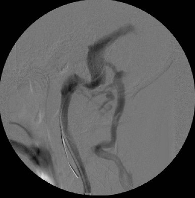
Images 1a, 1b (R-IJV, lateral): Is the vein to the right of the Sigmoid sinus the Right VV?
Image 3a (R-IJV, front): I think the blue arrow is the transverse dural sinus.
Images 2 and 3b (L-IJV, front): Any guess as to what the "squiggles" are? Vertebrals? Collaterals? Plexus veins?
Image 4 (L-IJV, lateral): Again (see Image 1a, 1b), is the vein to the right of the Sigmoid sinus the Left VV?
Do you have any post-valvuloplasty images showing if the VVs stopped "lighting up" with dye, or would that necessitate DrS having to go back into the SS to release more dye and use more radiation? Is it safe to believe that the VVs won't fill with dye anymore once the lower IJV valvuloplasty is completed?
~~~~~~~~~~
Cece, you set the bar very high with your searching abilities.
The composite image showing all those veins is fantastic. I was always unsure where exactly the lumbar veins are, and seeing them in this image clears up my confusion. I'm still unsure of where the renal veins are? I agree that the vein to the left of the Azygos is the Hemiazygos vein.
Here is a link to a great PowerPoint presentation that shows the dural sinuses and the bottom of the skull in all their glory (the d/l might be a bit slow). One can really see how vulnerable all the vessels and nerves are to shifts in the skull:
www.similima.com/ppt/anatomy/anat-cranial-fossa.ppt
Thanks, Cece! Great job!!
Posted: Thu Jul 21, 2011 4:11 pm
by Cece
Any questions I actually know how to answer?


I don't have any post-valvuloplasty images of the vertebral veins. From looking at the images, the right jugular is the healthier one, so the way the vertebral veins look on those two images might be the way the left side should look afterwards.
I'm just playing around when I search, usually. You never know what'll turn up. These were from a google image search of "vertebral veins" because I wanted to make sure I was correctly identifying the vertebral veins in my images above. I really like seeing the veins all the way from azygos arch to iliac.
Posted: Thu Jul 21, 2011 5:10 pm
by jgalt2009
Cece wrote:There's another labelled azygos vein lower down, what is that about? Is it the hemiazygos? Or is still the azygos?
According to Netter's Atlas, the arch of the azygos is about 2 vertebrae above the accessory hemiazygous, and about 3.5 above the hemiazygos. I think I can make out about 4 vertebrae between the arch and where the vein in question traverses posteriorly (assuming this is a right lateral view). But it seems to be an anterior view (note annotations of left and right illiacs), so if it is an anterior view, the "? vein" is moving laterally in the wrong direction.
I think this is a mirror image of an anterior view, and the annotations for left and right are juxtaposed. If so, the "question-mark" vein is also mis-marked and it is in fact, the hemiazygos.
But then again, what do I know?
Posted: Thu Jul 21, 2011 7:57 pm
by Cece
We are seeing the arch of the azygos in the direction we normally see it. It is not flipped.... So if we knew what view that is, we'd know what view this is.
It does kinda hurt the brain, doesn't it?
Posted: Thu Jul 21, 2011 8:18 pm
by Cece
HappyPoet wrote:CeImages 1a, 1b (R-IJV, lateral): Is the vein to the right of the Sigmoid sinus the Right VV?
I think so.
Image 3a (R-IJV, front): I think the blue arrow is the transverse dural sinus.
That sounds right
Images 2 and 3b (L-IJV, front): Any guess as to what the "squiggles" are? Vertebrals? Collaterals? Plexus veins?
I don't know. They are the same squiggle veins that are on my original image before my procedure in February:
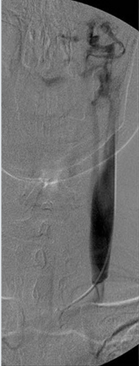
Image 4 (L-IJV, lateral): Again (see Image 1a, 1b), is the vein to the right of the Sigmoid sinus the Left VV?
I think so. What else could it be? My VVs are on the large size too.
Do you have any post-valvuloplasty images showing if the VVs stopped "lighting up" with dye, or would that necessitate DrS having to go back into the SS to release more dye and use more radiation?
I believe it would have required him to go back up into the sinus or near it.
Is it safe to believe that the VVs won't fill with dye anymore once the lower IJV valvuloplasty is completed?
I don't know. My verts are well-developed, for which I am grateful, they've served me well. I wonder if they would still take more than average just because they're there and they can? Or would the flow of the jugular win out? I think I have large verts on the other side too so I'll stick with my answer that the left side would, post-venoplasty, look more like the right side.
I'm still unsure of where the renal veins are?
I am not sure where they are in this image.
Here is a link to a great PowerPoint presentation that shows the dural sinuses and the bottom of the skull in all their glory (the d/l might be a bit slow). One can really see how vulnerable all the vessels and nerves are to shifts in the skull:
www.similima.com/ppt/anatomy/anat-cranial-fossa.ppt
Thanks for the link! I will have to watch it tomorrow, I got defeated partway through the first of Dr. Tucker's youtube videos....did you know that two waves going in the opposite direction, when they meet, it'll combine the pressure of both waves?
Posted: Thu Jul 21, 2011 8:46 pm
by Cece
Cece wrote:Ok, no changes in my numbness in my left for 8 days post-procedure and then in a matter of hours it melts away entirely.
This is a numbness that comes and goes but it does not go in this fashion, except once before, after my first venoplasty. I am sufficiently awed.
This morning, when I woke up, the first thing I realized was that my arm felt odd and the second thing I realized was that it felt odd because the numbness is still gone.
Other improvements specifically since this second procedure are that colors are brightened, my breath control is easier when reading out loud or drinking water, and my vision is sharper when it comes to making out lines, like the edges of buildings.
These are all improvements that I had the first time around. I was aware of losing the breath control and I was aware when the numbness came back but I wasn't aware of the colors getting less bright or any vision reduction. Even as these improvements were diminishing, other improvements like my foot drop and heat intolerance happened after that. Someone explain that one to me....
Emotionally I went through a dip where I was mad that I've had to live with subpar vision and subpar energy and, y'know, MS when others were seeing these sorts of colors all along. And I was sad because I am expecting restenosis this time, since it happened before, and who knows if it'll be my right jugular too. But, the first is me getting upset about something that is past and the second is me getting upset about something in the future. When I focus on right now, right now is unexpectedly great.