Page 4 of 4

Re: Another Important New BNAC CCSVI Study

Posted: Thu Nov 17, 2011 9:13 pm
by MSBOB
There are cases where trauma causes MS. Usually the MS is highly progressive with trauma.
(Editing, beacause i looked it up. It was a long standing assumption that Ms is caused by head teams, but there is little evidence)

I naturally meant neurological impacts of the vascular, autoregulatory and autoimmune defects.

I meant the white blood cells don't pass the Bbb unless they have a purpose, like fighting an infection, cleaning up debris, or repairing. Otherwise, they shouldn't be in the cns. Pml is caused by blocking BBB too much, leaving the brain vulnerable to opportunistic infections. Viruses don't need the space that white blood cells need to pass through the BBB. What is interesting, is that viruses pass through like a micronutrient, but cause the same hormones to be released that dilate the veins. This can leave the BBB permeable for an extended timeframe.

When the white blood cells pass through, they first adhere to the vessel wall, and "eat" through. They release hormones that cause the veins to dilate. Then other hormones like protein enzymes break a path through for the white blood cell. That is part of inflammation. That is the natural process. Tysabri interferes with the adhesion hormones, if i remember correct.

If the white blood cells don't clean the debris, it may just sit there and scar up. I don't quite know, but i think it doesn't get cleaned up. That is ok because brain cells live for 75 years or more, they don't need to regenerate as often as other less protected cells. There is usually not much to clean up. Waste like urea is small and passes freely through the BBB and is carried away in the blood. By debris, i mean dead cells.

Re: Another Important New BNAC CCSVI Study

Posted: Fri Nov 18, 2011 8:11 am
by Cece
MSBOB wrote:That is ok because brain cells live for 75 years or more, they don't need to regenerate as often as other less protected cells.
I think this is the problem: our brain cells are not as protected as other people's brain cells.
I would like my brain cells to be protected from myelin-sensitized t-cells, inflammation, hypoxia, venous congestion, poor CSF flow, starvation, toxic build-up, whacks to the head, viruses, bacteria, and wallerian degeneration. Not a comprehensive list but if we take care of all these conditions that would be an excellent start.

The model of blood-brain barrier breakdown due to inflammation is interesting but we also have a model of blood-brain breakdown due to abnormal shear stress conditions due to reflux. The two models are not mutually exclusive.
Image
"Promotion of migration" is due to the increase in adhesion molecules.

Re: Another Important New BNAC CCSVI Study

Posted: Fri Nov 18, 2011 9:03 am
by ttucker3
Cece

Two pictures are worth two thousand words. Are you able to provide a reference for the pictures? You might be interested in knowing that a lot of computational work has been done on shear stress associated with stenoses in the arteries, but I can't find any related to stenoses in the neck or head veins. This computational work predicts reversing positive and negative blood flow in one pulse cycle (as observed in MRI flow quantification measurements by Haacke et al.)

(For examples see http://www.sciencedirect.com/science/ar ... 9073900997 or

http://virtual2002.tau.ac.il/users/www/ ... enosis.pdf or

http://www.menet.umn.edu/~bett0099/8390_1.pdf

Trev. Tucker

Re: Another Important New BNAC CCSVI Study

Posted: Fri Nov 18, 2011 9:25 am
by Cece
I shouldn't have left off the attribution. Here it is:
from this article: http://www.nejm.org/doi/full/10.1056/NEJMra055289
here's the image but it's too small here: http://www.nejm.org/na102/home/ACS/publ ... 289_f4.gif

Re: Another Important New BNAC CCSVI Study

Posted: Fri Nov 18, 2011 9:42 am
by MSBOB
I would like my brain cells to be protected from myelin-sensitized t-cells, inflammation, hypoxia, venous congestion, poor CSF flow, starvation, toxic build-up, whacks to the head, viruses, bacteria, and wallerian degeneration. Not a comprehensive list but if we take care of all these conditions that would be an excellent start.
That pretty much sums up my views on it, too. I am glad that we have all lightened up a little. I do find all of thi interesting.

Re: Another Important New BNAC CCSVI Study

Posted: Fri Nov 18, 2011 1:04 pm
by Cece
It does not surprise me that there has not been models done of the head and neck veins. Such an under-researched area.
I will have to read the linked articles slowly, as I am poorly acquainted with both newtonian and nonnewtonian fluid dynamics, and Reynolds numbers, and vortex shedding.

http://virtual2002.tau.ac.il/users/www/ ... enosis.pdf
X-ray Angiography is error-prone when the vessel is noncircular, because it uses a projection view of the geometry.
Dr. Cumming showed us an azygous that looked stenotic by angiography, but nonstenotic by IVUS, because of the oval (noncircular) shape of the vein.
Experimental studies of stenotic vessels have typically focused
on the flow downstream of the stenosis, particularly on the disturbance
produced by the stenosis and whether the resulting flow is
turbulent, not on the mechanisms of plaque rupture.
In CCSVI, what we are interested in is the effect of the stenosis on the upstream flow. Downstream has been virtually ignored.

Here's an entirely different article, on downstream effects:
http://www.sciencedirect.com/science/ar ... 8404002084
Venous Flow Restriction: The Role of Vein Wall Motion in Venous Admixture

S Raju , , G Cruse, M Berry, S Owen, E.F Meydrech, P.N Neglen

River Oaks Hospital and University of Mississippi Medical Center, Jackson, MS, USA

Accepted 21 April 2004. Available online 7 June 2004.

Abstract

Objectives. There are wide differences in flow between vascular beds at rest, even more during stress. The hydrodynamic energy (Energy grade line or EGL) of venous outflows must also vary considerably between vascular beds. We explored the mechanism of venous admixture of differing energy flows using a mechanical model.

Materials and methods. The model simulated two venous flows coalescing at a venous junction and then flowing through collapsible venous pumps. Flow rates and pressures were monitored when the venous pumps were full (steady state) and when they were compressed and allowed to refill inducing wall motion (pump flow).

Results. With increasing EGL differences between two coalescing venous flows, reduction or cessation (venous flow restriction) of the weaker flow occurred during steady state; higher base EGL of both flows ameliorated venous flow restriction and lower base EGL the opposite. Outflow obstruction favoured venous flow restriction. Pump action in the vicinity of the venous junction abolished venous flow restriction and maximized both venous flows.

Conclusion. The model suggests a pivotal role for vein wall motion in venous admixture and regional perfusion. Observations in the model are explained on the basis of network flow principles and collapsible tube mechanics.
This one discusses outflow obstructions and veins, in a mechanical model. Dr. Raju and Dr. Neglen are experts in May Thurner syndrome. The base of the jugular, where valvular obstructions are, flows into the subclavian so looking at subsequent admixture might be possibly relevant. My limited understanding of this article is that the weaker flow coming from the occluded jugular would be further weakened by competition for flow in the faster-moving subclavian, further reducing outflow from the jugular. The pump would be the thoracic pump. Vein wall motion is greatly affected by elasticity of the vein, and females have less elasticity than males.

Ok, that's a thought on downstream flow.

Back to the first article....
Morphological details of diseased vessels, such as surface irregularity and stenosis curvature, have been shown to have important effects on the blood flow.
If a plaque is like a curved speed bump on the artery wall, wouldn't a fixed valve leaflet be more like a straight-out road block? If curvature matters, I would expect these sorts of stenoses to have a greater effect on flow.
Both pressure and velocity distributions are similar for all three
Reynolds numbers, with magnitudes of both pressure and velocity
increasing with Re. Figure 5 shows the contours of total pressure
~static plus dynamic pressure! for this range of Reynolds numbers.
A large pressure force acts on the wall just upstream of the stenosis
throat in the internal carotid artery.
There's a large pressure effect, just upstream. Interesting.
At diastole, vortex shedding
is observed from the shoulder of the carotid sinus, far upstream of
the most severe occlusion. The vortex that has formed at t50.230
seconds has detached by t50.290 seconds. This shed vortex is
carried into the sinus, where it dissipates. Another vortex has
formed at the shoulder by t50.50 seconds. This would indicate a
shedding frequency of 3.7 Hz, or a Strouhal number of 0.84 to
1.0.
ok, vortex shedding must be flow or pressure effects upstream or downstream of the stenosis itself. I don't want vortex shedding in my dural sinuses or capillaries of the brain.
The assumption of Newtonian
behavior may break down when the shear rate is low
(;0.1 s21!, for example in small arteries and downstream of
stenoses. In some diseased conditions, including atherosclerosis,
blood exhibits some non-Newtonian behavior @38,39#. The effects
of non-Newtonian behavior on flows in stenotic vessels are first
considered for axisymmetric 50 percent stenoses
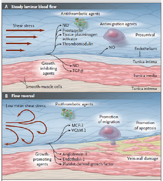
here we get to what's relevant to the shear stress diagram that I posted. What happens when the shear stress drops? We're also reading about arteries, which are high flow, which might not be directly applicable to veins, which are low flow.
The advanced lesion experiences a dramatically different flow
field from that of a healthy artery or one in the early stages of
atherogenesis. Fluid shear stress in undiseased arteries ~comparable
in size to the common carotid artery! is on the order of
1–2 N/m2, while endothelial cells in the throat of a tight stenosis
may experience shear stresses greater than 30 N/m2. As
Fry observed, shear stresses in this range may severely damage
endothelial cells, or even strip them from the vessel wall.
Tensile wall stresses, distributed evenly around the circumference
of a healthy vessel, are likely to be focused at the most vulnerable
shoulder regions of a diseased vessel @43#. ~The ‘‘shoulder regions’’
contain transitions from normal to diseased artery wall.!
This is discussing high shear stress, and the reason why plaques often rupture upstream of the point of maximum stenosis, and not at the maximum stenosis itself.

Gosh this is interesting. If the highest pressure is just upstream of the stenosis, then those of us with lower jugular stenoses may have been better off than those with high jugular stenoses, who may have a more complicated situation with the pressure being highest just upstream of that high jugular stenosis at an area much closer to the dural sinuses and the brain.
A k-v model such as that of
Wilcox, also given high marks by Patel et al., may be more
suitable than k-e models for flows in stenotic vessels.
k-v, not k-e
Our vessels are definitely stenotic.

My understanding of shear stress has only been of the effects of low shear stress, as seen in the diagram I linked. But this article talked about high shear stress existing at the site the stenosis itself, which makes sense, if flow velocity increases past the stenosis. The shear stress can be high enough to tear endothelial cells right off? Perhaps this is only true in high flow arteries, not low flow veins.

I am sure I did not understand nearly as much as I could have of this article, if I had the background, but it seems relevant to apply such mathematical concepts to CCSVI. I would like to see just what effect my bilateral stenoses (77% and 99%) had on the flow and pressure points upstream. An IR once suggested to a TiMSer who had a fully occluded vein that they might be better off with a full occlusion than with a partial occlusion. This seems nonintuitive, and obviously more outflow will be obstructed with a full occlusion than with a partial one, but it is possible that the full occlusion results in less reflux and less focal hypertension than a partial occlusion would. A mathematical model might help answer that question.

Re: Another Important New BNAC CCSVI Study

Posted: Fri Nov 18, 2011 1:04 pm
by Cece
:oops: Now that's a long post. It was a complex article....

Re: Another Important New BNAC CCSVI Study

Posted: Sun Nov 20, 2011 12:06 pm
by MSBOB
I am surprised that blood thinners have not shown much impact on MS. I know that from as far back as the 50s, doctors have hypothesized microembolism and thrombosis may contribute to the pathology of MS. Trials with blood thinners didn't seem to make a difference. Then later doctors discovered that turbulent blood flow and shear stress cause the release of endothelin. That also is shown in the diagram cece posted. Perhaps blood thinners don't tackle the problem.

My main concern with ccsvi treatment is that it may be undoing the body's natural work in its effort to protect the brain. I know that it seems like the opposite of what the IRs are saying, but if ccsvi is not congenital, than perhaps it is a beneficial response. Or it could have been a beneficial response that got caught in a feedback loop and became detrimental. Those are interesting questions. I think the neurologists may need to posit a countertheory of how MS causes ccsvi.

Re: Another Important New BNAC CCSVI Study

Posted: Mon Oct 15, 2012 10:28 am
by Cece
http://lib.bioinfo.pl/pmid:17703296
Parenchymal abnormalities associated with developmental venous anomalies.

Diego San Millán Ruíz, Jacqueline Delavelle, Hasan Yilmaz, Philippe Gailloud, Enrico Piovan, Alberto Bertramello, Francesca Pizzini, Daniel Rüfenacht
INTRODUCTION: To report a retrospective series of 84 cerebral developmental venous anomalies (DVAs), focusing on associated parenchymal abnormalities within the drainage territory of the DVA. METHODS: DVAs were identified during routine diagnostic radiological work-up based on magnetic resonance imaging (MRI)(60 cases), computed tomography (CT)(62 cases) or both (36 cases). Regional parenchymal modifications within the drainage territory of the DVA, such as cortical or subcortical atrophy, white matter density or signal alterations, dystrophic calcifications, presence of haemorrhage or a cavernous-like vascular malformation (CVM), were noted. A stenosis of the collecting vein of the DVA was also sought for. RESULTS: Brain abnormalities within the drainage territory of a DVA were encountered in 65.4% of the cases. Locoregional brain atrophy occurred in 29.7% of the cases, followed by white matter lesions in 28.3% of MRI investigations and 19.3% of CT investigations, CVMs in 13.3% of MRI investigations and dystrophic calcification in 9.6% of CT investigations. An intracranial haemorrhage possibly related to a DVA occurred in 2.4% cases, and a stenosis on the collecting vein was documented in 13.1% of cases. Parenchymal abnormalities were identified for all DVA sizes. CONCLUSION: Brain parenchymal abnormalities were associated with DVAs in close to two thirds of the cases evaluated. These abnormalities are thought to occur secondarily, likely during post-natal life, as a result of chronic venous hypertension. Outflow obstruction, progressive thickening of the walls of the DVA and their morphological organization into a venous convergence zone are thought to contribute to the development of venous hypertension in DVA.
This is not CCSVI research, but may shed more light on the effect of venous abnormalities including outflow obstruction on the parenchyma of the brain.

Re: Another Important New BNAC CCSVI Study

Posted: Tue Oct 16, 2012 6:46 am
by 1eye
cheerleader wrote:
Cece wrote:The argument that MS might cause CCSVI tends to be made by those who consider CCSVI to be a stricture of the vein itself and not as intraluminal abnormalities.

Is this suggesting that the CCSVI is causing the reduced metabolism and morphological changes in the brain?
I have the full paper, and the posits are that the MS disease creates hypometabolism of brain tissue and this leads to less blood flow and CCSVI, or CCSVI strictures create hypoperfusion and reduced visability of vasculature....
but Cece makes a GREAT point.
In studies, like the one from the Cleveland Clinic, announced today---intraluminal defects are found in the veins of pwMS at much higher rates than normals. These are not "strictures" or "stenosis" created by hypovolemia or inflammation---these intraluminal defects are truncular venous malformations, as we see in other congenital venous disease.
onward!
cheer
Look, let's connect the dots, here, shall we? Since "MS" doctors don't want to.

In no particular order:

dot 1. Cleveland study finds more intraluminal defects in "MS".
dot 2. Beggs, et al find higher hydraulic resistance to blood flow in CCSVI.
dot 3. Buffalo find reduced venous vasculature in "MS".
dot 4. There is reduced perfusion in "MS", starting, big surprise, before CCSVI/"MS" symptoms.
dot 5. Buffalo finds higher prevalence of CCSVI in "MS". Another big surprise.
dot 6. Buffalo finds more severity over time in CCSVI. This is a well-known feature of "MS".
dot 7. Buffalo find more collateral veins in necks of "MS" patients.

Please feel free to join in on the chorus at any time. I think the list is longer.

How many dots do we have to connect? The theory of CCSVI is not just a theory any more. It is supported by multiple independent and otherwise unrelated statements about both "MS" and CCSVI.

The tack of not acknowledging CCSVI exists, or that it has no sensible means of causing the symptoms of "MS", simply will not wash any more. Any more. Now, the people who are "against" this theory are resorting to imbecilic statements about the safety of angioplasty, in a foolish attempt to plug multiple holes at once, and somehow slow this progress at all costs.

At the same time big, big guns are being aimed at people who have used stents. Interesting that this is not anywhere close to happening in Poland, where stents are even more likely to be used. If you want to rake in profits in open court, using nothing but fear and deep pockets, California is more likely to allow you to. Stents or CCSVI angioplasties have not been used by Michael Dake in four years. Many more procedures, many more stents, no easier targets with as much to lose. As far as I am concerned, that makes the defendants even more heroic.

I for one, am not willing to waste more than nominal time on these studies or diatribes or press reports or bureaucratic pronouncements, which attempt to throw some kind, any kind, of cold water on CCSVI. I predict that the Saskatchewan trial and Brave Dreams will both show that angioplasty is not only safe but effective. I predict that the study run by the "MS" mafia in Montreal and BC will find some way to be negative about angioplasty. These are not difficult things to foresee. But sense is sense. The theory is getting stronger every day.

If anybody wants to send me a PM requesting my identity, I will respond. I am not afraid of anyone knowing who I am. I am a CCSVI, formerly "MS" patient, and I have no money to speak of, no financial interest in anything I say, or "MS", or CCSVI.

Re: Another Important New BNAC CCSVI Study

Posted: Tue Oct 16, 2012 8:03 am
by Cece
1eye wrote:How many dots do we have to connect? The theory of CCSVI is not just a theory any more. It is supported by multiple independent and otherwise unrelated statements about both "MS" and CCSVI.
But reading the ECTRIMS abstracts alone would lead to the opposite conclusion.
I do not think the evidence for CCSVI is a slam dunk, as of yet.

Re: Another Important New BNAC CCSVI Study

Posted: Tue Oct 16, 2012 8:26 am
by 1eye
It's slam-dunk enough that I think further promotion of the inferior "autoimmune" theory is foolish and causing much injustice and financial hardship, to say nothing of prolonging a doomed enterprise or suffering on account of it.

The "autoimmune" theory had its roots in work on allergies. While I believe they exist and cause harm (both my wife and brother have them, and have both encountered anaphylactic shock -- I may even have also done so), I think trying to pin "MS" on anything like allergy (what is it EAE stands for, again?) is at this point ridiculous.

So what are we left with? Injections of drugs that sometimes cause death, indentured slavery and mute acceptance, or picking up whatever marbles are left, and going home?