Page 1 of 1

adhesion molecules

Posted: Sat Oct 22, 2011 7:56 pm
by Cece
http://physiologyonline.physiology.org/ ... 4/166.full
Image
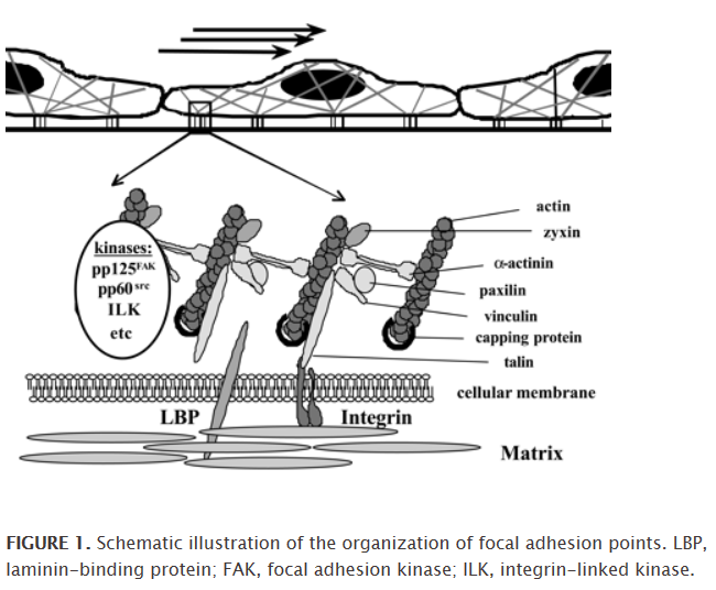
The image does very little to help me understand this. But if you have heard that reduced shear stress causes an increase in adhesion molecules, and you have thought, I know what shear stress is but what is an adhesion molecule, there it is. The arrows in the top image demonstrate shear stress, which should be one-way, but in pwCCSVI can be all turbulent and disturbed. The lined-up cells that look a bit like fried eggs, three in a row, still in the top image, are the endothelial cells. On the bottom of each endothelial cell are the dashed lines, as if those fried eggs had centipede feet, and those are the adhesion molecules.

Here's a quote on it:
Because of their special position, endothelial cells are ideally suited for sensing changes in blood flow velocity, which cause differences in shear stress. These changes in shear stress are directly responsible for numerous cellular responses, like release of autacoids such as nitric oxide, prostaglandins, and the endothelial-derived hyperpolarizing factor, which are all necessary for rapid adaptations in vessel diameter. Beside these fast and acute reactions, there are also known long-term responses that result, for example, in altered gene expression. According to a widely accepted hypothesis, shear stress is sensed by endothelial cells in so-called focal adhesion sites (Fig. 1) (9). These are structures in which clustered matrix receptors meet the cytoskeleton via certain linker molecules. Changes in their composition and/or cellular tension lead subsequently to the activation of kinases, which in turn initiate appropriate signaling cascades for altered gene expression.
It doesn't make me think, eureka, I understand! But it is useful to know that there are complex effects from disturbed blood flow.

edit: to be clear, the specific place where these endothelial cells and adhesion sites are being affected by the disruptions in shear stress caused by CCSVI is in the blood brain barrier, which is the endothelium or wall of the capillaries of the brain. In the autoimmune theory of MS, there was never an explanation for why the blood brain barrier was weakened, although it was known that it was. In CCSVI, we have an explanation.

Re: adhesion molecules

Posted: Sun Oct 23, 2011 8:22 am
by Cece
http://www.news-medical.net/health/What ... phage.aspx
When a leukocyte enters damaged tissue through the endothelium of a blood vessel (a process known as the leukocyte extravasation), it undergoes a series of changes to become a macrophage. Monocytes are attracted to a damaged site by chemical substances through chemotaxis, triggered by a range of stimuli including damaged cells, pathogens and cytokines released by macrophages already at the site. At some sites such as the testis, macrophages have been shown to populate the organ through proliferation.

Unlike short-lived neutrophils, macrophages survive longer in the body up to a maximum of several months.
A second preclinical study demonstrated that a signaling pathway for Copaxone(R) deactivated white blood cells called macrophages that induce inflammation and autoimmune response.

"Data have indicated that activated damaging macrophages may contribute to axonal loss in MS, and that the deactivation of these macrophages may be a therapeutic goal of treatment," said lead study author, Nicolas Molnarfi, Researcher Neuroimmunologist, Department of Neurology and Program in Immunology, University of California, San Francisco, California. "These data showed that Copaxone(R) deactivated specific macrophages, elucidating a potential mechanism for the impact of Copaxone(R) treatment."
http://www.msrc.co.uk/index.cfm/fuseact ... ageid/1767

I've thought the gradual improvements are odd. Why did I experience better heat tolerance and improvement in foot drop three months after my procedure? Neurological healing is one good explanation. But if macrophages survive in the body for several months, it could be a die-off of the pre-treatment macrophages, which might be sensitized to the myelin after they were invited in through the adhesion sites to clean up the damaged cells.

Re: adhesion molecules

Posted: Sun Oct 23, 2011 9:38 am
by Jugular
Improved motor neurological function following CCSVI treatment is difficult to explain. Improvement in cogfog or fatigue, these are relatively easy, but foot drop - not so much. I expect that we won't be getting much help from neurologists anytime soon, given the Ostrich reflex the bulk of them have about this issue. "Why explain something that isn't happening" I expect they'd say.

One good explanation that 1eye came up with was that brain plasticity becomes impaired as MS progresses. Improved drainage of the saturated environment might enable the brain to get back to adapting and improvising through new neural pathways.

In this recent study the authors suggest that RR MS can be explained by plasticity. In other words, the wounds don't go away, the brain just loses its ability to work around them.

http://www.mstrust.org.uk/professionals ... 008_05.jsp

Myself, I think that remyelination and nerve regeneration are also possible, though difficult.

Re: adhesion molecules

Posted: Sun Oct 23, 2011 9:44 am
by Cece
Jugular wrote:One good explanation that 1eye came up with was that brain plasticity becomes impaired as MS progresses. Improved drainage of the saturated environment might enable the brain to get back to adapting and improvising through new neural pathways.
Nerve cells would be all around healthier, with better oxygen, better glucose supply, and better waste removal. Makes sense to me that this would improve plasticity. I like it.

Re: adhesion molecules

Posted: Tue Oct 25, 2011 4:22 pm
by Cece
www.ncbi.nlm.nih.gov/pubmed/12909305
Brain Res Bull. 2003 Aug 15;61(3):357-64.

Adhesion molecules and matrix metalloproteinases in Multiple Sclerosis: effects induced by Interferon-beta.

Avolio C, Giuliani F, Liuzzi GM, Ruggieri M, Paolicelli D, Riccio P, Livrea P, Trojano M.

Neurology Unit, University of Foggia, Foggia, Italy. avolio@neurol.uniba.it

Abstract

In Multiple Sclerosis (MS) pathology, early inflammation involves leukocyte migration across the blood-brain barrier (BBB) within the central nervous system. In this process, adhesion molecules (AMs), both membrane-bound and soluble-circulating forms, and matrix metalloproteinases (MMPs) certainly play a regulatory role. In MS, recombinant Interferon-beta (rIFNbeta) is effective in reducing gadolinium contrast-enhancing lesions on magnetic resonance imaging and this suggests that it may reduce BBB damage or even restore its integrity by different mechanisms that include interference with both AM and MMP pathways. This review will highlight the effects induced by rIFNbeta, both in vitro and in vivo, on cell-bound and soluble forms of AMs and on MMPs.
Neurologists have heard of adhesion molecules too, and see them as playing a role in early inflammation in MS.