This Is MS Multiple Sclerosis Knowledge & Support Community
Welcome to This is MS, the leading forum for Multiple Sclerosis research and support. Join our friendly community of patients, caregivers, and researchers celebrating over 20 years of delivering hope through knowledge. https://www.thisisms.com/forum/
Wall shear stress:the missing step for Tcell transmigration?
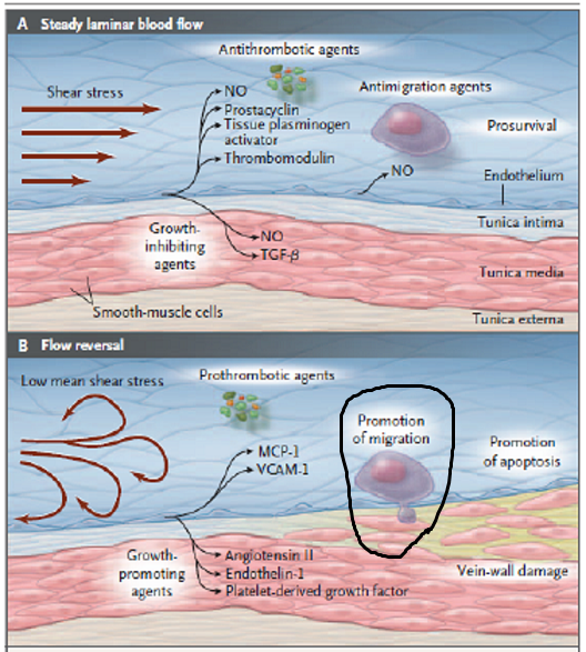
In CCSVI, shear stress is reduced. Here it is showing (I think) that shear stress pushes t-cells through the endothelium. We do not want T-cells crossing the endothelium of the capillaries of the brain, aka the blood brain barrier. A reduction of shear stress would in theory be good, except for the increase in adhesion molecules caused by the reduced shear stress. Adhesion molecules are the little "handholds" that the t-cell is grabbing onto before it crosses.
Re: Wall shear stress:the missing step for Tcell transmigrat
Posted: Wed Mar 28, 2012 2:39 pm
by David1949
This reminds me of the Daflon 500 video
regarding the manner in which leukocytes roll along the endothelium and then penetrate it.
Watch at about 2:20 into the video.
Re: Wall shear stress:the missing step for Tcell transmigrat
Posted: Wed Mar 28, 2012 3:01 pm
by Cece
there's always this image for discussions on shear stress!
I haven't watched that video yet, thanks Dave
Re: Wall shear stress:the missing step for Tcell transmigrat
Posted: Wed Mar 28, 2012 6:18 pm
by HappyPoet
There's also this great article and real-time video of T-cells crossing the BBB: http://www.physorg.com/news176652011.html Watching the video makes me cringe at the thought of what's been crawling around in my brain tissue.
Re: Wall shear stress:the missing step for Tcell transmigrat
Posted: Thu Mar 29, 2012 9:05 am
by 1eye
I think Cece's Nature reference implies that shear stress itself is the force which pushes the leukocytes through. Note that the article says that the stress is at a minimum at the vessel wall. It is associated with laminar flow, and is at a maximum at the centre of the vessel. Therefore the second diagram more accurately reflects the lack, or reduction. of shear stress as being the situation that allows unwanted transmigration due to the reduced force of low shear stress, caused by a low laminar velocity when reflux or stenosis occurrs. That makes more sense to me, as the presence of infection is likely to cause signalling molecules to pass into the bloodstream, attracting immune cells. When the signalling is achieved, perhaps the leucocyte expresses something in response, which opens the endothelium.
The laminar flow is perpendicular to the endothelium. If it is too strong, the leukocyte gets swept away. But at the endothelium, if VCAM is present, be rolled along, or stick in place, or transmigrate. If the reflux or stenosis causes not enough laminar force, or too much VCAM, it is too sticky to roll, and perhaps falls through, or starts expressing the opening "something" molecules.
I think the BBB is porous because of the stretching effect of increased venous pressure, which makes the size of the gaps in the endothelial sieve (tight junctions) become larger (less tight), letting unwanted molecules (iron? other metals? oxidants? leukocytes?) through.
Re: Wall shear stress:the missing step for Tcell transmigrat
Posted: Thu Mar 29, 2012 11:13 am
by munchkin
Would low blood pressure affect sheer stress?
It seems to me that low blood pressure would indicate slower flow and according to one of the IRs many of his patients have low blood pressure.
Re: Wall shear stress:the missing step for Tcell transmigrat
Posted: Thu Mar 29, 2012 1:32 pm
by 1eye
Blood pressure is typically not measured in a very thorough way. It is measured at the arm, with a cuff and a stethoscope, not inside the head. It is measured by listening to the sound. Because venous pressure is so low by comparison with arterial, I am thinking (possibly wrongly) that venous pressure is not measured at all with the cuff on the arm. The BBB is subject to arterial pressure on the arterial side, and venous pressure (much lower in every case) on the outflow side.
Ohm's law with fluids says that pressure = flow * flow resistance. That results in a pressure drop across the stenosis or narrowing. So if that is in the neck, then venous pressure, even if it were measured by an arm cuff, or some kind of plethysmographic method, would be higher on the brain side of the obstruction. I doubt that is what is referred to by the IR.
Would low blood pressure affect sheer stress?
It seems to me that low blood pressure would indicate slower flow and according to one of the IRs many of his patients have low blood pressure.
Pressure (not rate) should be lower post-obstruction, due to its resistance to flow. A flow measurement on either side of the stenosis should show that only pressure is affected, not flow rate; on either side of the stenosis, flow rate should be the same.
There are only two sources of cerebral blood pressure: muscles (heart and others) and gravity. Gravity is a major factor in drainage of areas which are physically above the heart, because the pressure due to other muscles pumping is low in comparison, due to pressure losses in the capillary beds and venules.
The spinal veins drain upward, when you are upright and they are below the heart. That may be why the azygus figures in CCSVI. You are in double jeopardy if the jugulars are blocked (brain drains sideways when prone) and the azygus is also blocked (spine and ribcage drain upward -- via azygus -- when upright. Parts are below heart).
That said, venous pressure lowered more than normal by a stenosis, would affect the laminar-ness (not the rate, except globally).
Re: Wall shear stress:the missing step for Tcell transmigrat
Posted: Fri Mar 30, 2012 1:08 pm
by 1eye
The laminar flow is perpendicular to the endothelium.
Did I say that? I meant to say parallel. However, the diagram shows how the forces can be parabolic, and have a component at a different angle.
Re: Wall shear stress:the missing step for Tcell transmigrat
Posted: Fri Mar 30, 2012 1:38 pm
by 1eye
I just thought: if the stenoses in the internal jugular are causing a pressure increase upstream in the brain, well gravity stays the same. Neck muscles probably don't do much pumping, and the only other source of increased pressure, needed to compensate for the stenosis's increased resistance, is the heart. So is MS progression happening because the heart is aging, or for some reason losing its ability to sustain the increased pressure which would be necessary to have the same flow rate as before? Perhaps the person, for whatever reason, cannot continue to add more collateral veins, so the person just stops having remissions. The heart driven pressure component cannot increase forever, and the stenosis gets worse of its own accord, because during the increased transit time, the blood loses more oxygen and glucose to keep the brain alive longer. Some experimental results contradict that, but I need verification of those. It's as if there is a minimum threshold of flow rate needed to keep the drains healthy, and beyond that, problems can't be compensated for, so they start to only cause more problems.
Re: Wall shear stress:the missing step for Tcell transmigrat
Posted: Fri Mar 30, 2012 1:43 pm
by Cece
1eye wrote:
The laminar flow is perpendicular to the endothelium.
Did I say that? I meant to say parallel. However, the diagram shows how the forces can be parabolic, and have a component at a different angle.
1eye, you lost me at parabolic...
What does it mean to have a component at a different angle?
Re: Wall shear stress:the missing step for Tcell transmigrat
Posted: Fri Mar 30, 2012 2:52 pm
by 1eye
mistake
Re: Wall shear stress:the missing step for Tcell transmigrat
Posted: Fri Mar 30, 2012 2:55 pm
by 1eye
A force which appears to act in one direction, can be thought of as the vector sum of two or more simultaneous forces acting in different directions. A curved line can be thought of as a series of points very close to each other.
In the bloodstream example, there is more friction (drag?) on the blood at the vein wall than there is in the middle, so if there were T-cells in a straight line from one vessel wall to the opposite wall, the blood flow would push them forward with more force in the centre, and they would end up in a parbola, rather than a straight line.
"In small venules, flow has been calculated as laminar shear stress with a parabolic velocity profile (minimum velocity at the vessel wall, maximum at the center line of the vessel)."
Maybe at junctions where the venules branch to smaller ones,the drag becomes very high, the VCAM accumulates there too, and so the lesions are just piles of dead T-cells which went through there and left a smear. (Takes hat off mouth. ).
Re: Wall shear stress:the missing step for Tcell transmigrat
Posted: Wed Apr 11, 2012 3:44 pm
by 1eye
OK,, if anyone is still interested in this: where are these low shear stresses occurring, that would affect their transmigration through the BBB? I surmise that they are above the stenosis, in the veins of the brain. Maybe the shear stress is least at the BBB; maybe it is least at the venule/ventricle/lesion site. It is perhaps the point of least shear stress that is the farthest point into the brain that refluxing venous blood reaches, which might also form part of the venous blood-brain-barrier, or VBBB.
Does refluxed blood ever cross the BBB the wrong way? Or are the capillary beds in the brain one-way-only?
Is this also where the leukocytes get across? Or is it at the lesion sites? I think there might've been something about that in the article about the T-cells creeping, the one with the film of T-cells...
In fact, wouldn't the least shear, and the greatest resistance, and the lowest velocity, of blood for which gravity is a significant part of the force moving it, be on the venous side of the capillary beds in the highest point above sea level as it were (depending on posture)?
Re: Wall shear stress:the missing step for Tcell transmigrat
Posted: Wed Apr 11, 2012 4:22 pm
by Cece
The effects on the brain would have to be taking place at the blood-brain barrier, which is the endothelium wall of the capillaries of the brain.
If blood flow is slowed down, there might be stasis in these capillaries. Or reflux. The low shear stress would increase the adhesion molecules, which would pull leukocytes across, which would munch away on whatever damaged/dying neurons and glial cells they find. There would also be diapedesis which is leakage of red blood cells across the blood brain barrier, causing iron-poisoning of the neurons, and giving the leukocytes more to munch on. I am not sure about your last statement, as I don't know where the least shear or greatest resistance or lowest velocity would be. The flow entering into the capillaries would be at high pressure, as the arteries and arterioles are, so there would be some push forward, wouldn't there? And then the veins are more of a passive receiving system.
I don't think we know yet what role CCSVI plays in MS lesion formation. If the lesions are fibrin cuff, as seen in CVI of the legs, then that has nothing to do with the leukocytes. If the lesions are inflammatory, which I would believe to be true, then they are occurring at areas where the blood-brain barrier has been breached.
Re: Wall shear stress:the missing step for Tcell transmigrat
Posted: Thu Apr 12, 2012 5:39 am
by 1eye
OK, so lets say the BBB is any endothelium in the brain or spinal cord. Is the point of the breach known to be in capillary beds, or at venule/ventricular lesion sites? I think I read Dr. Sclafani saying that there is something different at spots where there is an aneurysm or a dead-end or something. I think what we're finding, by the shape of the lesions, is that they occur at venule branch junctions, and appear to come from the place where the blood must go in one branch or the other and change direction, which would reduce shear, and increase turbulence. The closer to the apex of the junction, the more turbulence. Perhaps that is where the most adherence is, and leaks occur at the same time as the transmigration. (Or are caused by it?)
I guess what I'm trying to figure out is: is it right in the capillary beds, or not, that most of the transmigration occurs, or is it at lesion sites? Because the highest point in the blood's gravitational trajectory, if it is in capillary beds, would be the point of lowest velocity. That might be in different organs, depending on posture.
Does which side you get disabled on, depend on which side of your body you lie on? Because I've lately been lying on my right side more often, to give my heart a rest (based on what Dr. Oz said, which must be right because I saw it on TV.).