CAREFUL APPLICATION OF THE RECOMMENDED PROTOCOLS IMPROVE REPRODUCIBILITY AND PERMIT COMPARISON AMONG CCSVI STUDIES.
Paolo Zamboni and Mirko Tessari
Vascular Disease Center, University of Ferrara, Ferrara, Italy.
Dear Editor,
We read with interest the article published by Rodger and Co-Authors
"Evidence against the Involvement of Chronic Cerebrospinal Venous Abnormalities in Multiple Sclerosis. A Case-Control Study" (Plos One August 2013, Volume 8, Issue 8, e72495).
As correctly reported, in 2009 we described the presence of chronic cerebrospinal venous insufficiency (CCSVI) in patients with multiple sclerosis (MS), proven by color Doppler ultrasonography (CDUS) and confirmed by catheter venography.1
The results obtained by the Canadian researchers are exactly the antipodes of what we found, since they were unable to demonstrate any venous flow abnormalities in the MS patients investigated both with CDUS and MRV. We briefly discuss below, why the methodology adopted by the Authors may lead to a so strong discrepancy in comparing results.
i)
Regarding CDUS methodology, we were very surprised that the Authors failed to use the updated methodology recently recommended by an international consensus in order to improve the reproducibility of the CDUS protocol.2
The only meta analysis of all reports from 2005 till June 2011 demonstrated a strong prevalence of CCSVI in MS, but with marked heterogenicity among studies.3
To avoid this and to make the studies more comparable after June 2011, seven international scientific societies developed a technically detailed protocol, not cited yet.2,4
ii)
Contrary to the recommended protocol, we were impressed in the Rodger study by the absence of any M-mode analysis for investigating the criterion #3.2
CDUS M-mode is indispensable to detect intraluminal obstacles and fixed valve leaflets, which represents the majority of CCSVI venous abnormalities.4,5
In figure 1 the valve motility of a normal subject is well apparent as compared by a patient with CCSVI and MS. This represents an intraluminal obstacle leading to flow blockages and/or bidirectional flow depicted in the figure 2, and seen by several Authors, but, unfortunately, never detected in the survey reported by the Authors.1-5
iii)
Regarding MRV methodology we were again surprised by the focus of the investigation in the upper and mid region of the neck, where significant differences in jugular flow rate were never detected in CCSVI condition. To the contrary, several reports measured significant restriction of the jugular flow rate, increased flow through the collaterals, and extraluminal stenosis in the lower portion of the neck, exactly where Rodger et al. omitted to perform any assessment.6-1
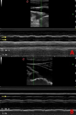
Figure 1.
A:
Two mobile valve leaflets evidenced in the M-mode trace by the yellow arrows. The lumen is never occupied and both leaflets appears well saddled to the jugular vein wall.
B:
A fixed monocusp valve in a CCSVI case, where the yellow arrow indicate the occupancy of the center of the lumen. This constitutes an intraluminal obstacle generating the flow abnormalities represented in the lower panel of figure 2.
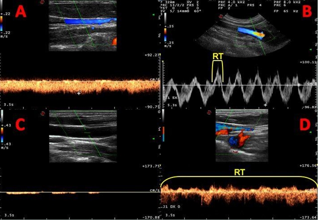
Figure 2.
Upper panels: normal flow traces in the internal jugular vein;
Lower panels: flow traces in CCSVI. In A a mono directional flow downward the chest (negative wave) is well apparent, as compared to the flow absence in a
CCSVI case depicted in C.
In B the flow in the valvular region is bidirectional for a reflux time (RT) < the cut-off 0.88 sec., whereas in CCSVI case, represented in D, the RT is > of
the cut-off (3.5 sec).
REFERENCES
1.
Zamboni P, Galeotti R, Menegatti E, Malagoni AM, Tacconi G et al. Chronic cerebrospinal venous insufficiency in patients with multiple sclerosis. J Neurol Neurosurg Psychiatry. 2009;80:392-399.
2.
Laupacis A, Lillie E, Dueck A, Straus S, Perrier L et al. Association between chronic cerebrospinal venous insufficiency and multiple sclerosis: a meta-analysis. CMAJ. 2011;8;183.
3.
Zamboni P, Morovic S, Menegatti E, Viselner G, Nicolaides AN. and the Intersocieties Faculty. Screening for chronic cerebrospinal venous insufficiency (CCSVI) using ultrasound. Recommendations for a protocol. Int Angiol. 2011;30:1-2.
4.
Zamboni, Paolo, Menegatti E, Occhionorelli S, Salvi F. "The controversy on chronic cerebrospinal venous insufficiency." Veins and Lymphatics 2.2 (2013): e14
5.
Zivadinov R, Ramanathan M, Dolic K, Marr K, Karmon Y, et al. Chronic cerebrospinal venous insufficiency in multiple sclerosis: diagnostic, pathogenetic, clinical and treatment perspectives. Expert Rev Neurother. 2011;11:1277-94.
6.
McTaggart RA, Fischbein NJ, Elkins CJ, Hsiao A, Cutalo MJ, et al Extracranial venous drainage patterns in patients with multiple sclerosis and healthy controls. AJNR Am J Neuroradiol. 2012;33:1615-20.
7.
Dolic K, Marr K, Valnarov V, Dwyer MG, Carl E, et al Sensitivity and specificity for screening of chronic cerebrospinal venous insufficiency using a multimodal non-invasivezimaging approach in patients with multiple sclerosis. Funct Neurol. 2011;26:205-14.
8.
Dolic K, Marr K, Valnarov V, Dwyer MG, Carl E et al Intra- and extraluminal structural and functional venous anomalies in multiple sclerosis, as evidenced by 2 noninvasive imaging techniques. AJNR Am J Neuroradiol. 2012;33:16-23.
9.
Feng W, Utriainen D, Trifan G, Elias S, Sethi S, et al.. Characteristics of flow through the internal jugular veins at cervical C2/C3 and C5/C6 levels for multiple sclerosis patients using MR phase contrast imaging. Neurol Res. 2012;34:802-9.
10.
Rahman MT, Sethi SK, Utriainen DT, Hewett JJ, Haacke EM. A comparative study of magnetic resonance venography techniques for the evaluation of the internal jugular veins in multiple sclerosis patients. Magn Reson Imaging. 2013 Jul 11. [Epub ahead of print].
11.
Utriainen D, Feng W, Elias S, Latif Z, Hubbard D, et al. Using magnetic resonance imaging as a means to study chronic cerebral spinal venous insufficiency in multiple sclerosis patients. Tech Vasc Interv Radiol. 2012;15:101-12.
12.
Feng W, Utriainen D, Trifan G, Sethi S, Hubbard D, et al. Quantitative flow measurements in the internal jugular veins of multiple sclerosis patients using magnetic resonance imaging. Rev Recent Clin Trials. 2012;7:117-26.
13.
Haacke EM, Feng W, Utriainen D, Trifan G, Wu Z, et al. Patients with multiple
sclerosis with structural venous abnormalities on MR imaging exhibit an abnormal flow distribution of the internal jugular veins. J Vasc Interv Radiol. 2012;23:60-8.
14.
Monti L, Menci E, Ulivelli M, Cerase A, Bartalini S, et al. Quantitative
Colour Doppler Sonography evaluation of cerebral venous outflow: a comparative study between patients with multiple sclerosis and controls. PLoS One. 2011;6:e25012.
15.
Zamboni P, Sisini F, Menegatti E, Taibi A, Malagoni AM, et al. An ultrasound model tocalculate the brain blood outflow through collateral vessels: a pilot study. BMC Neurol.2013;11;13:81.
Competing Interest: we have published results that are disputed in the commented article.1,15