Page 1 of 1

Impaired CSF pulsatility in healthy individuals

Posted: Tue Nov 12, 2013 8:35 am
by Cece
http://onlinelibrary.wiley.com/doi/10.1 ... 8/abstract
Aqueductal cerebrospinal fluid pulsatility in healthy individuals is affected by impaired cerebral venous outflow

Clive B. Beggs PhD1,*,
Christopher Magnano MS2,
Simon J. Shepherd PhD1,
Karen Marr RVT, RDMS2,
Vesela Valnarov MD2,
David Hojnacki MD3,
Niels Bergsland MS2,
Pavel Belov2,
Steven Grisafi BS2,
Michael G. Dwyer MS1,
Ellen Carl PhD1,
Bianca Weinstock-Guttman MD3,
Robert Zivadinov MD, PhD2,3

Article first published online: 8 NOV 2013

DOI: 10.1002/jmri.24468


Purpose


To investigate cerebrospinal fluid (CSF) dynamics in the aqueduct of Sylvius (AoS) in chronic cerebrospinal venous insufficiency (CCSVI)-positive and -negative healthy individuals using cine phase contrast imaging.


Materials and Methods


Fifty-one healthy individuals (32 CCSVI-negative and 19 age-matched CCSVI-positive subjects) were examined using Doppler sonography (DS). Diagnosis of CCSVI was established if subjects fulfilled ≥2 venous hemodynamic criteria on DS. CSF flow and velocity measures were quantified using a semiautomated method and compared with clinical and routine 3T MRI outcomes.


Results


CCSVI was associated with increased CSF pulsatility in the AoS. Net positive CSF flow was 32% greater in the CCSVI-positive group compared with the CCSVI-negative group (P = 0.008). This was accompanied by a 28% increase in the mean aqueductal characteristic signal (ie, the AoS cross-sectional area over the cardiac cycle) in the CCSVI-positive group compared with the CCSVI-negative group (P = 0.021).


Conclusion


CSF dynamics are altered in CCSVI-positive healthy individuals, as demonstrated by increased pulsatility. This is accompanied by enlargement of the AoS, suggesting that structural changes may be occurring in the brain parenchyma of CCSVI-positive healthy individuals.
This is new research from Dr. Beggs et al. They looked at healthy individuals, not MS patients. So this means studying CCSVI in itself, not CCSVI in multiple sclerosis. This is a good and necessary separation.

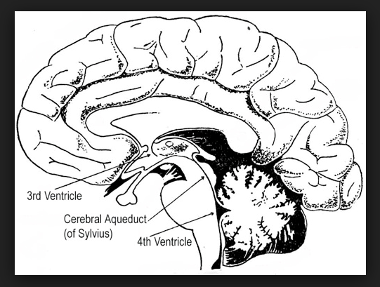
The healthy individuals with CCSVI had increased cerebrospinal fluid pulsatility in the AoS along with enlargement of the AoS. AoS is short for aqueduct of Sylvius. Not being a neurologist, I need a google image search to identify what the aqueduct of Sylvius is, and here we are:
http://www.nervous-system-diseases.com/ ... nosis.html
Image

I have to question the reliability of Doppler imaging to diagnose CCSVI since we've seen since such difficulty with this in other studies. But at face value, this research shows that CCSVI may be altering the dynamics of CSF and causing change to the structure of the parenchyma of the brain. That opens up a host of other questions, including if these changes are pathological or pathology-inducing in any way and if the changes reverse when CCSVI is treated.

This was good research to read on a cold Tuesday morning.

Re: Impaired CSF pulsatility in healthy individuals

Posted: Wed Nov 13, 2013 12:54 am
by frodo
Maybe the best part is to finally separate the existence of CCSVI from MS. Maybe now there will be less attacks to the CCSVI concept from the neuro community.

Maybe what we need now is a name for the "Impaired CSF pulsatility" and a doctor that test it on different populations

Re: Impaired CSF pulsatility in healthy individuals

Posted: Wed Nov 13, 2013 10:15 am
by 1eye
frodo wrote:Maybe the best part is to finally separate the existence of CCSVI from MS. Maybe now there will be less attacks to the CCSVI concept from the neuro community.

Maybe what we need now is a name for the "Impaired CSF pulsatility" and a doctor that test it on different populations
I dunno. The message to neurologists seems threatening to neurologists' turf. This is several outside specialties claiming to know more, and to be able to fix more, and be of more assistance to the sick in several areas where neurology has failed them. That puts the two (or more) sides in direct competition, for research dollars, patients, and air-time.

Meanwhile neurologists have staked out a position of skepticism that limits their options. If they say they don't need to do anything or be involved, then they can have no quarrel if someone else can do it. They are ceding real estate that they claim for themselves, by trying to discredit the work, at any price,

It's too bad, because they could have had a prime position of influence in some promising new area. By refusing to trust legitimate science, they have taken a position which is incompatible with future participation in that science.

I saw Michael J Fox on a talk show last night, He is continuing to work, in a high profile position, while his Parkinson's disease progresses. His face, its obviously mottled shades of bright red and pale areas, makes you think he must have some problem with circulation, some grave vascular condition.

Re: Impaired CSF pulsatility in healthy individuals

Posted: Wed Nov 13, 2013 1:57 pm
by vesta
1eye wrote:
frodo wrote:Maybe the best part is to finally separate the existence of CCSVI from MS. Maybe now there will be less attacks to the CCSVI concept from the neuro community.

Maybe what we need now is a name for the "Impaired CSF pulsatility" and a doctor that test it on different populations
I dunno. The message to neurologists seems threatening to neurologists' turf. This is several outside specialties claiming to know more, and to be able to fix more, and be of more assistance to the sick in several areas where neurology has failed them. That puts the two (or more) sides in direct competition, for research dollars, patients, and air-time.

Meanwhile neurologists have staked out a position of skepticism that limits their options. If they say they don't need to do anything or be involved, then they can have no quarrel if someone else can do it. They are ceding real estate that they claim for themselves, by trying to discredit the work, at any price,

It's too bad, because they could have had a prime position of influence in some promising new area. By refusing to trust legitimate science, they have taken a position which is incompatible with future participation in that science.

I saw Michael J Fox on a talk show last night, He is continuing to work, in a high profile position, while his Parkinson's disease progresses. His face, its obviously mottled shades of bright red and pale areas, makes you think he must have some problem with circulation, some grave vascular condition.
Thanks. Very well said.

Re: Impaired CSF pulsatility in healthy individuals

Posted: Thu Nov 14, 2013 2:24 am
by Robnl
You can undergo a CINE MRI, which makes a serie of photo's of CSF pulsation, combining the images makes a small movie

(In he FONAR study it was also done)

Re: Impaired CSF pulsatility in healthy individuals

Posted: Thu Nov 14, 2013 2:32 pm
by 1eye
Robnl wrote:You can undergo a CINE MRI, which makes a series of photo's of CSF pulsation, combining the images makes a small movie

(In he FONAR study it was also done)
I think they are not strictly photos, but that would be along the lines of what they did. I don't know what the "semi-automated method" was but if it wasn't that, it might verify this study if it were done in the way you suggest.

Interesting to me to see Dr.Zivadinov et al. working with Dr. Beggs I see this this as an excellent sign. Thank you researchers all!

He probably knows how to identify CCSVI by now.

Re: Impaired CSF pulsatility in healthy individuals

Posted: Thu Nov 14, 2013 9:12 pm
by Robnl
Hi. 1eye,

It' s called cinemagragraphic, to the way they made the first moties; make photos....
On June 15, 1878, under the sponsorship of Leland Stanford, Eadweard Muybridge successfully photographed a horse named "Sallie Gardner" in fast motion using a series of 24 stereoscopic cameras. The experiment took place on June 15 at the Palo Alto farm in California with the press present. The exercise was meant to determine whether a running horse ever had all four legs lifted off the ground at once. The cameras were arranged along a track parallel to the horse's, and each camera shutter was controlled by a trip wire which was triggered by the horse's hooves. They were 21 inches apart to cover the 20 feet taken by the horse stride, taking pictures at one thousandth of a second.[4]
http://en.wikipedia.org/wiki/History_of_film

:-D

Re: Impaired CSF pulsatility in healthy individuals

Posted: Fri Nov 15, 2013 10:17 am
by 1eye
Robnl wrote:You can undergo a CINE MRI, which makes a series of photos of CSF pulsation, combining the images makes a small movie
(In the FONAR study it was also done)
CSF flow and velocity measures were quantified using a semiautomated method and compared with clinical and routine 3T MRI outcomes.
It was remarkable what was done in early cinematography. The horse photography was synchronized, the same idea that cardiac-gated MRI applies electronically. But although film used to be used for MRI images, now I believe they are mostly electronic. That is all I meant
in saying they were not strictly photographic: No photons are necessary.

I believe MRI is done, like CAT scans, using a circling, spiraling sensor. However I was only referring to the fact that an MRI image measures magnetic and electromagnetic energy at radio frequencies, along with that which is generated by a large fast-moving magnet. This magnet must be swung around the subject, making a huge whacking sound when it is moved.

This is not from information I have read, but mainly I have just surmised it from what I have experienced when inside these machines. I believe the magnet to be huge because it makes so much noise. It is likely a combination of a real magnet, which would be heavy, and a coil conducting a large current. This combination makes an extremely strong magnetic field (measured in Teslas, which are a very large unit of magnetic force).

That kind of field strength also comes from the large electromagnets used to move heavy metal objects like junk cars, and also speeding trains levitated magnetically over a track, and also particles in giant accelerators, which are miles in a circle, and moved at speeds approaching that of light.

The signals from these magnets and coils can be detected by other coils, because a magnetic field can induce a current in a wire (there are none of these detecting coils in the junk-moving-type electromagnets). The signal generation and detection coils in these MRI machines use the same principles as those used in phonograph cartridges back in the 1980s (I still buy these once in a while).

----------------------------
As an aside, a story about one of my MRIs: The guy doing this MRI was talking to me afterward, so I mentioned to him that I had felt something crawling up my back while I had been in there. I had been wearing jeans and a hospital johnny-shirt. He lifted up the johnny-shirt and plucked several metal nuts off of my back.

I had been working earlier that day on my son’s little bicycle. These nuts were about 1/2 inch wide, and ¼ inch thick. One is told to remove all metal objects, glasses, jewelry etcetera, and it is put in a locker for you. They had told me my belt-buckle was OK, but I had emptied my pockets of change.

Unremembered were the four steel nuts in one of my back pockets. They had crawled up my back, stuck to my skin. I was heavier then, and my weight held them down.

The MRI guy said that I had been lucky that they had not gotten loose, because, for one thing, they would have damaged the MRI, flying around inside, but also because “they would have gone through you, like a bullet”. I was suitably thankful that they had not, and got an idea of the strength of these magnets.
----------------------------

Use of MRI slices can be made in 2 or 3 dimensions. That is how we can see the 3D images. Keeping the same locations of the scans, but allowing time to pass, adds the 4th dimension - the time aspect, making them cinematic. Synchronizing, or gating, with heartbeats , adds accuracy and removes extraneous information, like the strings tied to the horse’s hooves. I think this pulsatility study, like many others in this area of research, are landmarks.

I believe Dr. Zivadinov has a very good idea of how the MRI and Doppler tests compare to each other. Combined with Dr. Beggs’ and Dr. Zamboni’s work using tilt-tables and plethysmography, and Dr. Haacke’s MRI work, as well as BOLD MRI work, we are gaining a good understanding of hemodynamics and brain circulatory (blood and CSF) physiology. When their measurements meet the math and the physics, they are unassailable. I think they all deserve a round of Internet-applause.

Re: Impaired CSF pulsatility in healthy individuals

Posted: Fri Nov 15, 2013 10:40 am
by Robnl
It was remarkable what was done in early cinematography. The horse photography was synchronized, the same idea that cardiac-gated MRI applies electronically. But although film used to be used for MRI images, now I believe they are mostly electronic. That is all I meant
in saying they were not strictly photographic: No photons are necessary.
Think we talk about the same thing slightly different; ofcourse its electronic nowadays.
But they put the quickly taken, static, mri images next to each other....hey...a movie :mrgreen:

Did you see the fonar cine mri's of pulsating csf?

Re: Impaired CSF pulsatility in healthy individuals

Posted: Sat Nov 16, 2013 5:19 am
by Robnl


A video of dr damadian