Posted: Fri Jun 12, 2009 10:02 am
Why, that was a GREAT article well worth the read. 
Welcome to This is MS, the leading forum for Multiple Sclerosis research and support. Join our friendly community of patients, caregivers, and researchers celebrating over 20 years of delivering hope through knowledge.
https://www.thisisms.com/forum/
said the spider to the fly...Sharon wrote:what a tangled mess of webs you have.
I thought mine were low res, but the one from the linked site is REALLY low res. I doubt yours were that bad?Mrhodes40 wrote:I do not know anything at all about this subject at all but mine looked more like this...
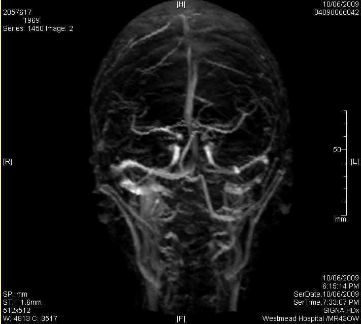
PeekaBoo wrote:Your doppler looks quite different than mine....I had no spaghetti but just 2 veins in the rear of my head
I am far from knowledgeable on this subject, however, my stab in the dark, with my eyes closed, is that the MR machine has a setting for what minimum diameter of vein to image, and below which size to ignore. I guess they used a standard setting, with the sensitivity turned all the way up, and hence the "cloudy" appearance because all the superficial veins were also imaged. Dr Dake knows he is looking at the larger veins, and so sets this control appropriately, where my guys used their standard settings. And if you forgot how this paragraph started, I don't know what I am talking about.cheerleader wrote:your pics look like the superficial/facial veins....Jeff's looked like the pics Marie linked to
whyRwehere wrote:I'm just guessing, but I think they weren't focused deep enough.
I think they were focused (for the little area they did) deep enough, as all the veins appear in the image (from all depths), my guess is that they just had the sensitivity turned up to high in the part of the computer system that interprets what comes back from the scanner, into an image. After reading the first few posts, I took to the images with a photo manipulation software, and if you alter the brightness and contrast of the image, it heads towards the "expected image'.PeekaBoo wrote:my thoughts exactly...maybe a higher strength 7 tesla?
ohh, stop, I'd blush, but it may affect my next scan.Cheerleader wrote:Cure, you handsome devil!
NO they were clear as a bell, but the thing that is different is that the jugulars were the lighted up thing not the little surface veins which were as if invisible, more like the linked picture.........Mrhodes40 wrote:
I do not know anything at all about this subject at all but mine looked more like this...
I thought mine were low res, but the one from the linked site is REALLY low res. I doubt yours were that bad?
peekaboo wrote:Your doppler looks quite different than mine....I had no spaghetti but just 2 veins in the rear of my head shaped like goggles and then of course the jugs...the contrast between black & white was much clearer...no mist
I recently had Dr Dake say he was happy to review the images that were taken, and comment on what was available. When preparing it to send to Dr Dake, I looked over the images on the disk, and found another sequence, that looked more like what others may of expected.mrhodes40 wrote:It looks different than mine as well. Mine seemed to have more of a deep vein view the bigger deeper veins and not so much of the nework on the skin which appears to be what is going on in your maybe?
...
On yours the jugs a look almost like a shadow instead of lighted up big and clear? I will be interested to hear What Dr D says about it
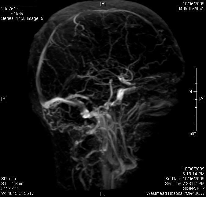
One thing I also noted, which I guess I never expected, is how asymetrical the insides of my head areDR Dake wrote:...Unfortunately, the study itself is somewhat noisy with a lot of arterial overlap that obscures resolution of the venous anatomy. I looked at every image of every sequence and I can say that the left sided venous drainage is asymmetrically diminutive compared to the right. It is difficult to pinpoint focal severe lesions rather it is more diffuse in nature. Bottom line, before doing anything else I would proceed to have a CT venogram from the aortic root up to the top of the head. This will clearly delineate the venous anatomy. It is much more operator independent that MR, but it does involve some radiation....
 I'm going to have another look at alex's shots. My brain (in the MRI's) is also lopped sided...