Page 1 of 2
If the pictures I've seen cause problems, then I'm dead!
Posted: Thu Oct 22, 2009 8:39 am
by beechwood297
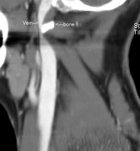
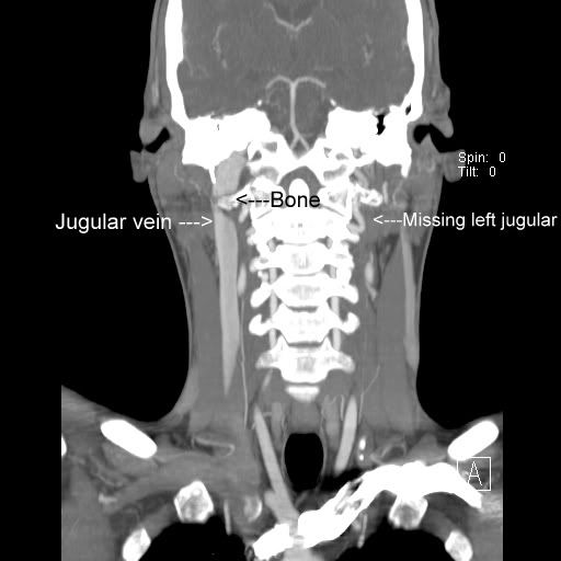
Three years of symptoms (cognitive difficulties, tinnitus, fatigue) and no answers. My scans show I only have one jugular instead of two, and the one I have is severely pinched. No answers from any Dr's so far, so maybe this can be a first step for me. Check these scans out. I hope this works ;)
Thanks for your interest,
Joel
Posted: Thu Oct 22, 2009 9:05 am
by HappyPoet
Oh my!!! You poor thing.
Those pictures have just got to make it into a textbook.
What test produced those amazing images?
I hope the pictures can put you on the road to good health. Keep us posted.
Thanks for sharing.
~HP
Posted: Thu Oct 22, 2009 9:06 am
by mormiles
Hi Joel and welcome! The good part of your only jugular looks really good, but yeah, that pinched part is an "eye-grabber." You're in the right place here. I hope you can get set-up soon for an appointment with Dr. Dake to fix it. Stanford is the really right place.
Posted: Thu Oct 22, 2009 9:45 am
by cheerleader
Welcome, Joel
Glad you visited us at Facebook. Welcome to TIMS. Who took these picks and why? Do you have an MS diagnosis? Do you have demyelinating lesions in your brain/spine? Wow...these are incredible images. I would set up a consult with Dr. Michael Dake at Stanford University.
MICHAEL D. DAKE, M.D.
Professor, Department of Cardiothoracic Surgery
Stanford University School of Medicine
Falk Cardiovascular Research Center
300 Pasteur Drive
Stanford, CA 94305-5407
t: 650.725.6407
f: 650.725.3846
e:
mddake@stanford.edu
The most interesting thing for me is to see how your bone is occluding the jugular vein. Certainly looks like a congenital situation-
please keep us posted!
cheer
Posted: Thu Oct 22, 2009 9:54 am
by whyRwehere
Oh dear,
this is what I am worried about for my husband. His doppler shows a missing jugular, but I am hoping that it is just pinched somewhere and no blood is flowing down it....but now you have realised my worst thought....So, what's the plan??
Posted: Thu Oct 22, 2009 9:58 am
by radeck
Hopefully that vertebrae extension can be removed! I'm very glad you haven't experienced worse symptoms so far. Looks like other blood routes are trying hard to do the job at least so far.
Posted: Thu Oct 22, 2009 10:07 am
by cheerleader
Joel-
Just saw on Facebook that you're from Michigan-
perhaps you should also give our friend Dr. Mark Haacke a call...
he is currently testing for CCSVI in Michigan, using SWI MRI (to see iron deposition/hypoxia in the brain) and MRV to study the veins. I know he'd be interested in your case-
The MRI Institute for Biomedical Research
440 East Ferry Street
Detroit, MI 48202
Email:
info.mrimaging@gmail.com
313-758-0065 Tel
313-758-0068 Fax
http://www.ms-mri.com/
collaborating with Dr. Haacke-
James Garbern
Dept. of Neurology, 8A University Health Center
4201 St. Antoine, Detroit, MI 48201, USA
(313) 745-4275
(313) 745-4468
j_garbern@wayne.edu
cheer
Posted: Thu Oct 22, 2009 10:16 am
by radeck
Looking at the image again, it seems like on the side where you have no jugular at all has the same bone sticking out. Could this have caused that jugular to be completely pinched of and wither away? In other words, I'm just offering an explanation of your situation that relies on only one congenital problem, those vertebrae extensions (if that's what we're looking at) on both sides. The other explanation would be that that second jugular was always missing, but seems less likely...
Posted: Thu Oct 22, 2009 4:23 pm
by beechwood297
HappyPoet wrote:Oh my!!! You poor thing.
Those pictures have just got to make it into a textbook.
What test produced those amazing images?
I hope the pictures can put you on the road to good health. Keep us posted.
Thanks for sharing.
~HP
CT scan with contrast was done because of a feeling of "pressure" in my head and base of skull. I also had (have) some difficulty swallowing along with a list including vision problems (floaters so bad I have a hard time reading) tinnitus, fatige, difficulty concentrating, hyper senstivity to sharp, sudden sounds, sleep apnea. No diagnosis, no mention of MS.
One Doctor will tell me "this is the type of thing people publish papers about" and another will say it is nothing and another says there is nothing that can be done about it- "veins don't like to be messed with" and such.
I had a vascular MRI done also. No answers. I'll try to post more scans if I can find the cd.
Posted: Mon Nov 02, 2009 11:55 am
by Sharon
Joel - the lower image showing the bone occluding the vein looks to me like the same image that is now on Dr. Haacke's website
http://www.ms-mri.com/index.php. Click on the left hand images -- go to case #3. Does Haacke have copies of your scans? Are you Case #3? Or, I wonder if Haacke has found another person similar to you.
Sharon
Posted: Mon Nov 02, 2009 12:04 pm
by cheerleader
Sharon wrote:Joel - the lower image showing the bone occluding the vein looks to me like the same image that is now on Dr. Haacke's website
http://www.ms-mri.com/index.php. Click on the left hand images -- go to case #3. Does Haacke have copies of your scans? Are you Case #3? Or, I wonder if Haacke has found another person similar to you.
Sharon
Sharon, I hooked up Joel with Dr. Haacke- and he's going to see him soon. That is his CT scan on the site.
Joel, you're famous

Now, let's get you feeling better!
cheer
Posted: Mon Nov 02, 2009 3:55 pm
by ozarkcanoer
beechwood... Your MRIs are astounding and amazing. I hope with all my might that when you see Dr. Haacke that it will be the first step on a journey to health for you. I will watch your posts with great interest.
Posted: Mon Nov 02, 2009 9:25 pm
by whyRwehere
With all due respect to Dr Haacke, but does he just make the images, or is he actually going to do any sort of procedure? Because although it is nice to have pictures...it's nicer to have some action on them!!
Posted: Tue Nov 03, 2009 5:02 am
by ozarkcanoer
No, Dr Haacke does NOT do the stenting procedures. But his 3D images are amazing and he is trying to develop a protocol for imaging on all people who have MS or are suspected of having MS.
I am participating in Dr. Haacke's study because I can drive to Detroit in 9 hours, because I may discover that I have CCSVI (Dr. Haacke does not do ultrasound... his disclaimer states that Zamboni's ultrasound may be necessary to discover all venous malformations). And because its free. But most of all I want to do something to help people with MS.
If I DO have venous problems that show up on his images, then I can approach a vascular surgeon in St. Louis to discuss stenting or other surgery. We have some excellent vascular surgeons here who may want to get in on the ground floor of CCSVI.
Posted: Tue Nov 03, 2009 6:51 am
by patientx
whyRwehere wrote:With all due respect to Dr Haacke, but does he just make the images, or is he actually going to do any sort of procedure? Because although it is nice to have pictures...it's nicer to have some action on them!!
Just for people that may not know, and I am not trying to minimize his credentials, but Dr. Haacke is not an medical doctor. He is a PHd in physics.