Page 1 of 1
Buffalo CTEVD Study MRV images
Posted: Sat Nov 28, 2009 7:57 am
by Dragonfly
I participated in the CTEVD study on Monday and Tuesday of this week, while I was home in Buffalo visiting family for Thanksgiving. I'm glad I set this up back in September, because they were swamped with phone calls on Monday after the CTV W5 program! It was a great experience, I had the dopplers, MRI/MRV, study questionnaire, even a mini neuropsych exam and a neuro exam from Dr. Bianca Weintock-Guttman, one of the PIs on the study.
So, as I think we all know, no MRI/MRV/doppler results are given to the participants (being a blinded study and all), but I was able to get a CD of my MRI/MRV images. I am going to bring the CD to work on Monday (I work at a medical university) to see what my boss the MD thinks (not a neurologist or vascular doc, though). However, I am curious to see what more knowledgeable people than myself think of these images. I really don't know what I'm looking at, and I'm having a hard time finding a "normal" MRV image online, so I have nothing to compare mine to. Any thoughts/opinions/comments are most welcome.
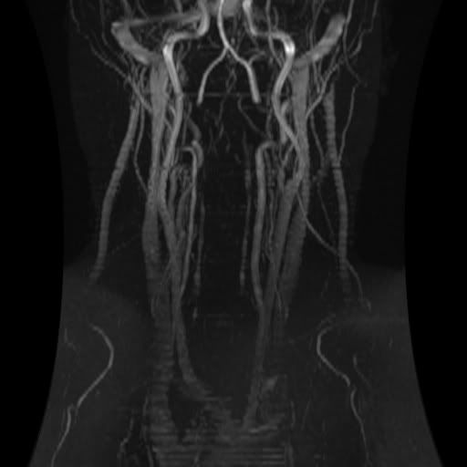
Without contrast:
With contrast:
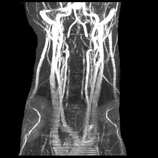
Looks funky to me in many respects, but I truly don't know what I'm looking at.
Posted: Sat Nov 28, 2009 8:29 am
by jimmylegs
neither do i but i agree re funky! it's pretty mixed up looking, got a nice spinning 3d one?

Posted: Sat Nov 28, 2009 10:02 pm
by jay123
Dragonfly - you have a PM-
MRV in buffalo
Posted: Sun Nov 29, 2009 7:09 am
by mickb
Hi Dragon -
You were smart to have set this up early. I get the MRI's this week at Buffalo but I was too late into the study to get an MRV. They said they only did the MRV's for the first 200 to get some correlation to the Doppler (which I got two weeks ago). I think having the MRV images will be important to move forward into any treatment. Now I have to figure out how I get the MRV. BTW, my understanding is the first "unblinding" is likely to occur very soon - in the first half of December.
Mick
Posted: Sun Nov 29, 2009 8:29 am
by Dragonfly
Mick, thanks for the info. When I was there earlier in the week, I asked about the first unblinding, and the research coordinator I spoke with said that it would occur sometime after they had enrolled 500 participants (I was #434). She thought that would happen by the end of the year, but I thought she said it would take some time to analyze the initial data? Who knows. They looked like they were up to their ears in work, though.
Rebecca
Re: Buffalo CTEVD Studyu MRV images
Posted: Mon Nov 30, 2009 12:25 am
by NHE
Hi Dragonfly,
I believe that Dr. Dake in Stanford uses 3-D image renderings to examine the veins as in some patients the veins can look fine from straight on but then are clearly flattened (or pancacked) when looked at from a 90° rotation. Do you have any way of viewing a 3-D rendering of the image files that Jacobs gave you?
NHE
Posted: Mon Nov 30, 2009 7:12 am
by Dragonfly
You can view the images in 3D, but I'm not really sure how to post that, or if anyone would be interested in looking at it. I was just kind of curious for some unofficial opinions.
Rebecca
Re: Buffalo CTEVD Studyu MRV images
Posted: Tue Dec 01, 2009 12:42 am
by NHE
Dragonfly wrote:You can view the images in 3D, but I'm not really sure how to post that, or if anyone would be interested in looking at it. I was just kind of curious for some unofficial opinions.
The utility you use to view the images in 3-D may have an option for exporting an image file of the current view. In addition, most things that can be displayed on your monitor can be captured using a screen capture. Press the Alt + Prt Scr (Print Screen) keys at the same time. This will copy the contents of the current window to the clipboard. You can paste it to an image file within any image editing application where it can be cropped and resized as you see fit. If you don't already have an image editing application, then you can download a free one from
http://www.gimp.org. This program has much of the same functionality as other programs such as Photoshop and Paint Shop Pro.
NHE
Posted: Tue Dec 01, 2009 1:31 am
by CureIous
If you'll accept layperson's opinion I'll shoot.
See the upside down wishbone doohicky in the middle? (my wife likes "doohicky" when I talk engines and stuff) , the veins immediately to the right and left of that are your IJV's if I am not mistaken. The one on the right (actually the left side of your body) does appear a bit suspicious from my untrained eye. Hopefully I left enough space here for the egg lol.
If you look at it a bit further down from my reference point (the upside down wishbone), where it narrows down a bit before widening, that would be what I would be interested in seeing from different angles.
Greater minds than I, greater minds...
Mark.
Posted: Tue Dec 01, 2009 1:56 am
by Algis
You might be interested in this; depending on how much you can deal with software:
http://www.sph.sc.edu/comd/rorden/dicom.html
Hope any will find it useful,
Algis
Posted: Tue Dec 01, 2009 8:43 am
by chrishasms
123
Thin ICE
Posted: Tue Dec 01, 2009 4:38 pm
by alphons
I am walking on thin ice here


I'm looking at the red dots.
anyone?
Posted: Tue Dec 01, 2009 4:42 pm
by CureIous
bingo.
Posted: Tue Dec 01, 2009 7:06 pm
by chrishasms
123
Posted: Wed Dec 02, 2009 9:00 am
by Dragonfly
Thank you to everyone who replied. I'm from Buffalo, I like to play armchair quarterback.

At this point, I'm more freaked out by the absent transverse sinus, way up at the tippy-top of the images above, above the wishbone doohickey on the right side. I have one one the left (though a little narrow), none at all on the right. Here's a better view (axial):
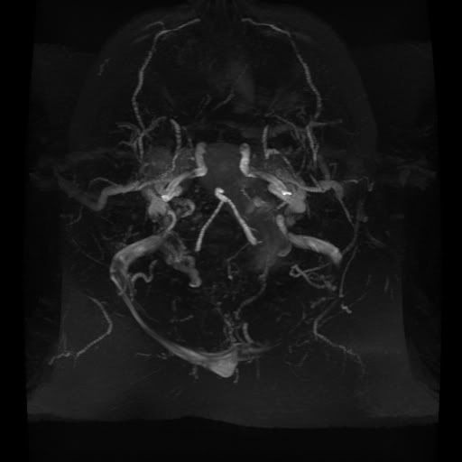
Then again, it's so high that I'm sure nothing could be done anyway. I just have to figure out who I can talk to about this. *Sigh*
Rebecca