Page 1 of 4
Hi all, Leetz asked me to post her MRV Pics
Posted: Wed Jan 06, 2010 8:37 pm
by CureIous
Posted: Thu Jan 07, 2010 7:08 am
by dialed_in
The (left?) IJV looks much smaller to my completely untrained eye. Is that just a normal one side dominant thing like being right handed?
Thanks for putting up all these pics in these different threads Mark, very interesting stuff. Do you have any posted of a non-ms'er system? I'm just wondering what a normal one would look like?
thanks
ETA: Ok, so I played around in photoshop a bit so that I could ask you some questions about these Mark. Hope you and Leetz don't mind.
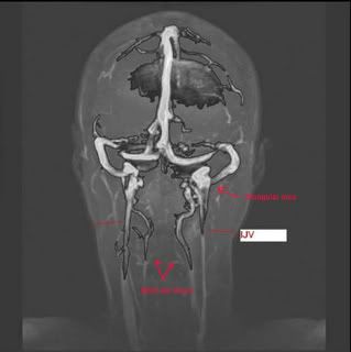
I can't remember what setting I clicked to get all that shading, but that's how photoshop sees it. The one thing I can't get over after looking at it for a while is the lack of symetry. Are all MRI/MRV images like this? I'm thinking that maybe if there were other slices that were taken closer and further from the origin you might see more symetry in them. So anyways, my questions:
The triangular area: Why does it look so different? Is it simply not going to look the symetrical to the other side because of the angle or depth at which the picture was taken? Sort of like what I was saying above?
What veins are the two in the middle?
I'm guessing that this picture was taken looking at Leetz's face. So right would be left and left would be right?
With that in mind her left IJV looks much thinner than the right. The left also has the triangular area which almost seems swollen in these pics, but that could be explained away by my questions above about slices and angles that the pictures were taken at.
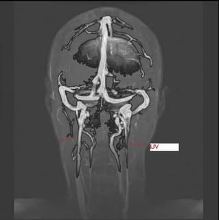
Ok, I was playing around again to figure out how I did that last picture. I used the magic wand to highlight the veins and then made a new layer. Then I replaced the colour on the layer to whatever photoshop had it defaulted too. I guess it was black because that's how all the shading came about. I really have no idea how photoshop does any of that, I'm really just messing around here. Here's another view with more of the veins selected:
Posted: Thu Jan 07, 2010 8:16 am
by Mechanicallyinclined
so that top pic where you have the ijv maked is a definite ijv too narrow correct?
Posted: Thu Jan 07, 2010 8:43 am
by annad
Has there been a stenosis diagnosis on leetz?
Posted: Thu Jan 07, 2010 8:52 am
by dialed_in
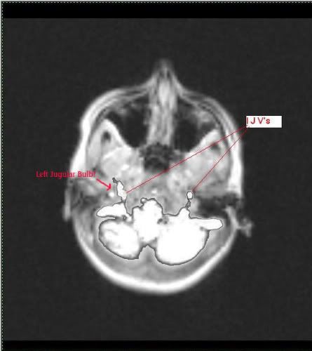
One more pic with a question about the slices again. This is the top down view, so easier to tell which is right and which is left. On the left the area is much larger and not a circular shape like the right side is. Is this just because of the slices? ie. would a slice lower down would look like a circle?
Is that the jugular bulb that we are seeing because it's higher up in her head than it is on the right side?
HELLO ALL...
Posted: Thu Jan 07, 2010 11:25 am
by leetz
THIS IS LEETZ...THANK YOU MARK!!!! WONDERFUL JOB...well they said no stenosis..lol...
#1--testing was done on a 1.5T by a TECH!!!!
#2 it was read by a neuro-radiologist(WHY???) these are vascular issues!!!
#3 NO DOPPLER
#4 i can see narrowing and my one IJV looks flattened and is not circular
#5 i am going to bring these to a vascular radiologist--and request to have a doppler done...
ANY SUGGESTIONS ON WHAT I SHOULD BE ASKING THE VASCULAR RADIOLOGIST?????
DOES
Posted: Thu Jan 07, 2010 9:09 pm
by leetz
does anyone see that down by the neck area the left IJV looks flat almost...not so much wide but flat???please tell me or do you think i am bugging out????
Posted: Thu Jan 07, 2010 9:24 pm
by dialed_in
Again, my eye is untrained, Mark would have a better idea after looking through his own so much. But in this picture:

The left(on the right side of the picture) looks tiny compared to the right (left side of the picture)
If you look at the size of the C shaped sections at the top, they are both the same size. Look lower down and the left thins right out while the right stays about the same size all the way down. That just looks wrong.
I have no idea if that's considered normal or not though?
Posted: Thu Jan 07, 2010 9:51 pm
by Mechanicallyinclined
I see a flat twisting in the right vein (left side of pic) . Left vein seems to be narrowing alot as it goes downward. (Right side of above pic) I'd like to be corrected on that if I'm wrong though.
Posted: Thu Jan 07, 2010 11:33 pm
by CureIous
Mechanicallyinclined wrote:I see a flat twisting in the right vein (left side of pic) . Left vein seems to be narrowing alot as it goes downward. (Right side of above pic) I'd like to be corrected on that if I'm wrong though.
Yup, that's what I saw on the right side, the twisting. The left needs hardly any explanation. I THINK those ones in the middle are either the vetebral veins, or the carotids, but although they seem too "twisty" to be carotids, if I was a betting man, I'd say carotids in the center.
Methinks these techs are looking for some type of "V'd" in pinched "really out there you can't miss it" stenosis, and when it doesn't come with a big arrow pointing to it, they err on the side of caution and make it a no-go.
Hopefully with Dr. Haacke's proto out there now the imaging centers will get up to speed, and I mean like yesterday.
Mark.
P.s. also on the left one, I'm getting the imression that it too is twisting, but we are looking at the side of it and it's wider looking from the side, but as a qualified journeyman uni0n trained pipefitter, I can assure you that flat pipes don't carry much fluids no matter how much pressure they are under....
misreading poor mrv
Posted: Fri Jan 08, 2010 4:53 am
by jak7ham9
I think that a lot of us are going to get lousy readings . I had a doppler done and my lef jugular was deemed to be much smaller greatly reduced flow some backwards flow visible. Doc diagnosed as stenois but tech was very vague. On my 1.5 tesla mrv reading again they had ststements that left jugular never wellseen much smaller in caliber not well seen froma hyperplastic left transverse into the inferior neck. These gguys (neuro radio) don't know how to read the reports also stated hard to see limited visability at skull base and upper neck becaue of turbulence. Hey I basically got the ccsvi diagnosis but the writing are really unclear beacause they don't know wht they are looking for. I think I need a 3 tesla reading by someone more knowledable. It is hard to do surgery without real knowledge. barbara
thank's to all...
Posted: Fri Jan 08, 2010 12:50 pm
by leetz
THANKS TO MY MS FAMILY....thank you soooo much everyone...i too saw everything that has been pointed out here...will visit with a vascular radiologist for 2nd opinion and will keep an updated post!!!!God Bless all of you!!!
LEETZ
Posted: Fri Jan 08, 2010 12:59 pm
by annad
All I can offer you is a cheer!!!!!
Go Leetz!!!!!!!!!!!!!!!!!
Crazy ride, isn't it??!!
best, best wishes!
a
annad
Posted: Fri Jan 08, 2010 1:04 pm
by leetz
THANK YOU...for your support it means so much!!!
LEETZ
Posted: Fri Jan 08, 2010 4:19 pm
by jimmylegs
hi guys can you shrink those images a bit to maintain the right page width? very interesting viewing

thx leetz


