DrSclafani answers some questions

A forum to discuss Chronic Cerebrospinal Venous Insufficiency and its relationship to Multiple Sclerosis.
User avatar
drsclafani
Family Elder
Posts: 3182
Joined: Fri Mar 12, 2010 3:00 pm
Location: Brooklyn, New York
Contact:

Post by drsclafani »

MikeInFlorida wrote:
drsclafani wrote:I have great suspicion that a stent will be necessary to treat such a stenosis due to chemotherapy stenosis.

Dr. Sclafani, this is disconcerting.
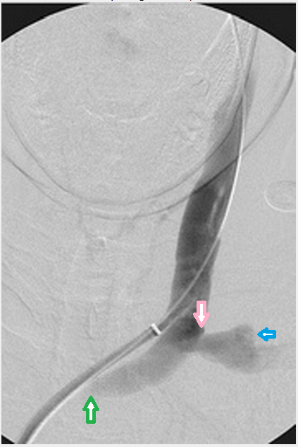
1. How bad was the stenosis in the innominate?
The stenosis was quite narrow in the innominate vein. The 3mm sheath completely obstructed the vein. But many stenoses are assymptomatic. for example in one report most of the stenoses and occlusions of dialysis related jugular stenoses were clinically occult. By this, i mean that the diagnosis was unrecognized by physical exams. there is usually no mention of neurological signs or fatigue, memory issues in the reports, which are usually described by radiologists.

In the gurley case reports on IJV and dural sinus thrombosis, an innominate stenosis was detected in one case (does figure 11 in the paper look familiar? :Melissa B. Gurley, Teresa S. King and Fong Y. Tsai (1996) Sigmoid Sinus Thrombosis Associated with Internal Jugular Venous Occlusion: Direct Thrombolytic Treatment. Journal of Endovascular Surgery: August 1996, Vol. 3, No. 3, pp. 306-314.).
2. What are the characteristics of a chemotherapy stenosis versus the typical stenoses you encounter?
In CCSVI encounter mostly valvular stenosis, occasional hypoplasia, infrequent and J2-J3 lesions, possibly due to inflammation and compressions. In chemo stenoses, this is intimal hyperplasia and scarring.
3. Specifically, do chemotherapy stenoses weaken the endothelial wall? Are they more delicate? More prone to thrombosis? More prone to elastic rebound?
chemotherapy stenoses are actually two types: one due to the cancers themselves. these are extrinsic. Catheter induced or chemotherapy induced stenosis are thickening of the endothelium, inflammatory scar and prone to elastic recoil. I think they are more likely to develop thrombosis but could not find the reference.

4. With respect to stenting, my recollection of the general tenor your historical posts is one of great reluctance to utilize stenting. Could you please elaborate on why a chemotherapy stenosis may require a stent?
The long term outlook for simple balloon angioplasty is not great. Most patients will require periodic dilatation. However intimal hyperplasia and compression of the stents are well known in this area. So I am inclined to see who well our patient tolerates this and a second angioplasty, and how well she responds to the angioplasty from her ccsvi symptoms. I am very comfortable with second look before making any decisions on this.
5. Do you believe that both the subclavian and innominate chemotherapy stenoses may require stenting?
possibly. time will tell. We certainly bit off quite a lot in the first treatments, didnt we?.

6. Would a stent placed in the innominate be riskier than one placed in the subclavian (specifically, more prone to travel, or more dangerous if it did travel)?

Both have risk but i would think that either can be managed by stents if necessary with small risk of migration. However because the stents are larger, migration is particularly "annoying"

7. Do any other options exist?
Surgical resection and graft interposition in the vein is an alternative. However comparisons between surgery and IR are bipolar. One study showed better results for surgery (against early experience with stenting). However a followup report from the same group showed similar results.
User avatar
drsclafani
Family Elder
Posts: 3182
Joined: Fri Mar 12, 2010 3:00 pm
Location: Brooklyn, New York
Contact:

Post by drsclafani »

MikeInFlorida wrote:Dr. Sclafani,

Here is an image of the IJV - subclavian junction from Gray's anatomy: Image

Here is a similar X-ray image of the patient:
Image

The junction point in the patient's X-ray looks nothing like the Gray's image. The subclavian seems to funnel down dramatically. The junction is neither the port origination point nor the termination point- it is somewhere in the middle of the port length.

1. Is this funneling actually there (or is it possibly a deceptive image?), and if so, is funneling normal (perhaps an artifact of the fact that the baloon is currently inflated)?
2. If the funnel is really there and is abnormal, would you consider this to be another stenosis?
3. If it is a stenosis, is it treatable?
Mike, great questions.
I think that funneling is real. This is confirmed by the collaterals that are seen, perhaps not on the image you posted.
BTW there is no balloon inflated on that image.
I do consider this a stenosis. and it was treated.
User avatar
drsclafani
Family Elder
Posts: 3182
Joined: Fri Mar 12, 2010 3:00 pm
Location: Brooklyn, New York
Contact:

Post by drsclafani »

Cece wrote:Image
I took the liberty of adding some arrows, to make sure we are discussing the same areas....

The blue arrow looks like a likely candidate for the subclavian stenosis, as lit by refluxing contrast. Just below the pink arrow is where you are seeing a funnel-like narrowing? The green arrow is where the contrast ends. This might be the innominate stenosis.

The jugular itself looks like a nice size.

I believe there is flow in the subclavian and innominate veins at all times, unlike the jugulars which collapse when upright, so that might be better for a stent if necessary. But let's hope the ballooning holds. What sort of follow-up imaging can be done to check on the innominate and subclavian? Is a doppler sufficient?
actually the pink arrow is what I thought was a stenosis and what was dilated. There were collaterals going around this area.
The green arrow is in the middle of the innominate vein which opacified very slowly because of the outflow obstruction.

I will followup with another venogram. Also perform a doppler trying to see below the clavicle. It might not work but worth the try.
User avatar
drsclafani
Family Elder
Posts: 3182
Joined: Fri Mar 12, 2010 3:00 pm
Location: Brooklyn, New York
Contact:

Post by drsclafani »

MikeInFlorida wrote: One other thing for the Doctor... I know that the catheter routing shown is SVC to innominate to IJV, but the section of catheter from the green arrow to the end of the sheath (assuming the end of the sheath is the white band) looks like it is outside the innominate. Is this a distortion of the image, or the result of the sheath blocking the contrast? If the lumen includes the area occupied by the catheter in the image, then the CSA of the innominate (between the green and pink arrows) looks substantial.
that is an illusion partially caused by inadequate opacification of the innominate vein.

the innominate vein is a big vein. The size of the vein does not reflect the narrowing of the stenosis which is relatively short and focal.
User avatar
drsclafani
Family Elder
Posts: 3182
Joined: Fri Mar 12, 2010 3:00 pm
Location: Brooklyn, New York
Contact:

Post by drsclafani »

Cece wrote:. Dr. Sclafani has said that he believes the stenosis was due to the puncture, which would be at the port site.
Allow me to clarify this. The site of the port is NOT the site of puncture into the vein. the puncture into the subclavian vein is going to be m ore central toward the right than the port. The port is placed in a convenient space not necessarily at the site of the puncture. We can only speculate until Mike posts a picture of a chest xray with the port and catheter in place. Then we can resolve this speculation.
User avatar
drsclafani
Family Elder
Posts: 3182
Joined: Fri Mar 12, 2010 3:00 pm
Location: Brooklyn, New York
Contact:

Post by drsclafani »

Cece wrote:
Plus there is still a whole RIJV to look at. And maybe an azygous or two. (With CCSVI, you never know....)

No pressure, Dr. Sclafani..... ;)
Maybe a little pressure.....
This case has generated a lot of interest!
patience my patient!
Still answering questions on this fascinoma
User avatar
drsclafani
Family Elder
Posts: 3182
Joined: Fri Mar 12, 2010 3:00 pm
Location: Brooklyn, New York
Contact:

Post by drsclafani »

HappyPoet wrote:Mike, did your wife's left arm ever swell before the venoplasty? If not, maybe collateral veins weren't needed; i.e., blood flow was slow but adequate enough for the body not to create collaterals. Thx.
the flow in the arm is well handled by collateral veins. The problems are that those collaterals are also important for cerebrospinal venous drainage. This is sort of like the ffect we have discussed with stenosis of the left iliac vein and the renal vein, although the cause is quite different.
Cece, your puff looks like it has a right angle in it with the point of your blue arrow pointing to the vertical line of the puff. I wonder if the puff is part of the port. Thx.
ignore the blue arrow
User avatar
drsclafani
Family Elder
Posts: 3182
Joined: Fri Mar 12, 2010 3:00 pm
Location: Brooklyn, New York
Contact:

Post by drsclafani »

Cece wrote:
MikeInFlorida wrote: Image
Looking at the image, do you see how the thyroid veins connect to the innominate vein? Thyroid veins may be recruited to serve as collaterals when jugulars are blocked. If the innominate is blocked too, depending on where the blockage is, it might prevent the thyroid veins from being effective collaterals.
on top of which the subclavian vein stenosis is creating collateral circuits between the thryocervical branches of the subclavian vein and the rest of the flow through the thyroid. In otherwords, competition for flow by both arm and brain.

really interesting case. I am glad there is so much interest in it!
User avatar
drsclafani
Family Elder
Posts: 3182
Joined: Fri Mar 12, 2010 3:00 pm
Location: Brooklyn, New York
Contact:

Post by drsclafani »

HappyPoet wrote:
Cece wrote:If the innominate is blocked too, depending on where the blockage is, it might prevent the thyroid veins from being effective collaterals.
I wonder how much of the arm's blood the thyroid veins can handle.
while you might not see them, there are many collateral veins for the arm, not just the thyroid. around the shoulder to the back, down the chest wall, . But also up the vertebral vein in some cases.
User avatar
drsclafani
Family Elder
Posts: 3182
Joined: Fri Mar 12, 2010 3:00 pm
Location: Brooklyn, New York
Contact:

Post by drsclafani »

MikeInFlorida wrote:
Cece wrote:
MikeInFlorida wrote: Image
Looking at the image, do you see how the thyroid veins connect to the innominate vein? Thyroid veins may be recruited to serve as collaterals when jugulars are blocked. If the innominate is blocked too, depending on where the blockage is, it might prevent the thyroid veins from being effective collaterals.
Your knowledge of anatomy is impressive. The thyroid collateral is a very good pickup.

I feel like I am in a game of "Clue", except I know a few of clues in advance (but for the most part, I am clueless). Since the good Doctor has already passed the ECD pre-op discussion, I guess I'll let you (and HappyPoet) in on this piece of 411: the pre-op ECD showed: either engorged thyroid collaterals (hopefully), or something else (best case, cysts, worse case, the big-C).

So here's my speculation on the good news and bad news: the good new is, you don't have thyroid cancer, and the thyroid is serving as an effective collateral. The bad new is, your innominate is pretty much blocked.
if the thyroid veins were effective, there would be no ccsvi and perhaps no hypothyroid disease.
User avatar
drsclafani
Family Elder
Posts: 3182
Joined: Fri Mar 12, 2010 3:00 pm
Location: Brooklyn, New York
Contact:

Post by drsclafani »

MikeInFlorida wrote:
HappyPoet wrote:Mike, does your wife show signs of thyroid dysfunction? Hyper or Hypo?
She does have hypothyroidism, and takes 50 mcg of generic Synthroid each day. She is scheduled to see an endocrinologist next week.
HappyPoet wrote:Do you know why the port has not been removed (sorry if I missed this)?
The port was removed 16 months ago. It was installed for the 18-month cytoxin series, then we left it in for an additional 20 months. It was left in place in case we decided to do another chemo series. After awhile, my wife wanted it out, so it was removed.
This was discussed earlier in the case but I wanted to share this reference, co-authored by my AAC partner, Kevin Sullivan.

remember this report, kevin?

CARDIOVASCULAR AND INTERVENTIONAL RADIOLOGY
Volume 26, Number 2, 123-127, DOI: 10.1007/s00270-002-2628-z
CLINICAL INVESTIGATIONS
Incidence of Central Vein Stenosis and Occlusion Following Upper Extremity PICC and Port Placement
Carin F. Gonsalves, David J. Eschelman, Kevin L. Sullivan, Nancy DuBois and Joseph Bonn
a 7% incidence of central vein stenosis or occlusion was found in patients with prior indwelling catheters and normal initial venograms. Catheter caliber showed no effect on the subsequent development of central vein abnormalities. Patients who developed new or worsened central vein stenosis or occlusion had significantly (p = 0.03) longer catheter dwell times than patients without central vein abnormalities. New central vein stenosis or occlusion occurred in 7% of patients following upper arm placement of venous access devices. Patients with longer catheter dwell time were more likely to develop central vein abnormalities. In order to preserve vascular access for dialysis fistulae and grafts and adhere to Dialysis Outcomes Quality Initiative guidelines, alternative venous access sites should be considered for patients with chronic renal insufficiency and end-stage renal disease.
While discussion is in patients with dialysis catheters, similar findings are noted with chemo, pacemaker and other IV access catheters.
Longer catheter dwell time most likely to develop central vein stenoses and thromboses.
User avatar
MikeInFlorida
Family Member
Posts: 31
Joined: Sun Aug 07, 2011 2:00 pm

Post by MikeInFlorida »

drsclafani wrote:In the gurley case reports on IJV and dural sinus thrombosis, an innominate stenosis was detected in one case (does figure 11 in the paper look familiar?

Image
I photoshopped a quick side-by-side for ease of comparing (Gurley case on the left, Dr. S's patient on the right). Even stenosed, my wife has prettier veins.

With respect to the risk of stent migration in subclavian versus the innominate, you said
DrSclafani wrote:Both have risk but i would think that either can be managed by stents if necessary with small risk of migration. However because the stents are larger, migration is particularly "annoying"
Annoying? Wouldn't migration into the SVC (then into rt. atrium) be potentially fatal?
Last edited by MikeInFlorida on Sun Aug 14, 2011 12:45 pm, edited 1 time in total.
User avatar
MikeInFlorida
Family Member
Posts: 31
Joined: Sun Aug 07, 2011 2:00 pm

Post by MikeInFlorida »

drsclafani wrote:Allow me to clarify this. The site of the port is NOT the site of puncture into the vein. the puncture into the subclavian vein is going to be more central toward the right than the port. The port is placed in a convenient space not necessarily at the site of the puncture. We can only speculate until Mike posts a picture of a chest xray with the port and catheter in place. Then we can resolve this speculation.
Thank you for clarifying. I assumed the subclavian vein access was just beneath the port access.

In an earlier post, you stated that the subclavian stenosis was likely the result of a vein puncture. Until now, I believed that to mean an injury due to an inadvertent puncture during the administration of the chemo. Now I believe that you were referring to the vein puncture that was necessary for the port tube to be inserted. Is this correct?

With respect to the site of the vein puncture for the port tube insertion, what mechanism is used to seal this site? Does it endothelialize? If so, the act of removing it seems to be prone to trauma.

I'll request the x-rays on Monday, and I'll post them as soon as I get them.
User avatar
drsclafani
Family Elder
Posts: 3182
Joined: Fri Mar 12, 2010 3:00 pm
Location: Brooklyn, New York
Contact:

Post by drsclafani »

MikeInFlorida wrote:
drsclafani wrote:In the gurley case reports on IJV and dural sinus thrombosis, an innominate stenosis was detected in one case (does figure 11 in the paper look familiar?

Image
I photoshopped a quick side-by-side for ease of comparing (Gurley case on the left, Dr. S's patient on the right). Even stenosed, my wife has prettier veins.

With respect to the risk of stent migration in subclavian versus the innominate, you said
DrSclafani wrote:Both have risk but i would think that either can be managed by stents if necessary with small risk of migration. However because the stents are larger, migration is particularly "annoying"
Annoying? Wouldn't migration into the SVC (then into rt. atrium) be potentially fatal?
Mike
if you notice the quotation marks, they are there for a reason. I was intending to have poetic understatement.

You have to know me better...


I would prefer to perform periodic dilatation than stenting in that area, however if it doesnt work, then stenting is on the table as is surgical repair. But data shows no significant difference on outcomes and one is far more invasive than the other.

Let's cross this bridge when we come to it.
User avatar
drsclafani
Family Elder
Posts: 3182
Joined: Fri Mar 12, 2010 3:00 pm
Location: Brooklyn, New York
Contact:

Post by drsclafani »

MikeInFlorida wrote:
drsclafani wrote:Allow me to clarify this. The site of the port is NOT the site of puncture into the vein. the puncture into the subclavian vein is going to be more central toward the right than the port. The port is placed in a convenient space not necessarily at the site of the puncture. We can only speculate until Mike posts a picture of a chest xray with the port and catheter in place. Then we can resolve this speculation.
Thank you for clarifying. I assumed the subclavian vein access was just beneath the port access.

In an earlier post, you stated that the subclavian stenosis was likely the result of a vein puncture. Until now, I believed that to mean an injury due to an inadvertent puncture during the administration of the chemo. Now I believe that you were referring to the vein puncture that was necessary for the port tube to be inserted. Is this correct?
Yes, not just the hole but the prolonged catheter insertion into this area
With respect to the site of the vein puncture for the port tube insertion, what mechanism is used to seal this site? Does it endothelialize? If so, the act of removing it seems to be prone to trauma.
A fibrous sleave extends from the catheter vein site and extends into the tissue surrounding the catheter back toward the port. It is possible that removing it can tear the venous tissue at the site of the entry but it is more likely that the healing process causes the stenosis.
I'll request the x-rays on Monday, and I'll post them as soon as I get them.
that will be very revealing.
Post Reply
  • Similar Topics
    Replies
    Views
    Last post

Return to “Chronic Cerebrospinal Venous Insufficiency (CCSVI)”