DrSclafani answers some questions

A forum to discuss Chronic Cerebrospinal Venous Insufficiency and its relationship to Multiple Sclerosis.
Cece
Family Elder
Posts: 9335
Joined: Mon Jan 04, 2010 3:00 pm
Contact:

Post by Cece »

drsclafani wrote:The entire vein was angioplastied with 8 mm balloons. (fig 7-8)The final film shows a residual stenosis which remained after several dilatations. (figure 9).

So now I will place my first stent in one of my patients.
With the vein having been hypoplastic and then if it were left with a high-up stenosis limiting blood flow, the likelihood of the vein clotting up is high. Maybe the stent prevents that outcome.
Figure 1 shows stenosis of the external jugular vein (circle).
figure 2 shows balloon angioplasty
figure 3 shows no residual stenosis in the left external jugular vein
Very nice! I'd expect external jugular vein stenoses to be along the lines of May Thurner or renal vein abnormalities, meaning no greater incidence of them in patients with MS then patients without MS but, if they are present, they may complicate the outflow in an already complicated outflow situation.

Very happy for the patients, respectively, for their foot drop improvements!!
User avatar
drsclafani
Family Elder
Posts: 3182
Joined: Fri Mar 12, 2010 3:00 pm
Location: Brooklyn, New York
Contact:

Post by drsclafani »

drsclafani wrote:
WeWillBeatMS wrote:Dr. Sclafani,

Are the external jugular veins also tested for CCSVI? I know that they have not proven to be a regular problem but what about for the people like myself who have lost hearing in one ear? And at times even my good ear rings and pops and feels plugged like my bad one does all of the time.

WeWillBeatMS
The external jugular drains the face and scalp. I do not study these veins and i do not think these veins are the first line of action, perhaps they take on a greater potential when the internal jugular cannot be opened or has clotted off.

On Saturday i treated two patients with footdrop and weakness of the lower extremity. Both had exciting reduction in foot drop within the first day. I am ever cautious to have too much excitement on early findings. the proof isin the durability of these improvements.

ONe of the patients turned into a tour de force and I would like to share with you some unusual techniques that will hopefully make a difference.
Image

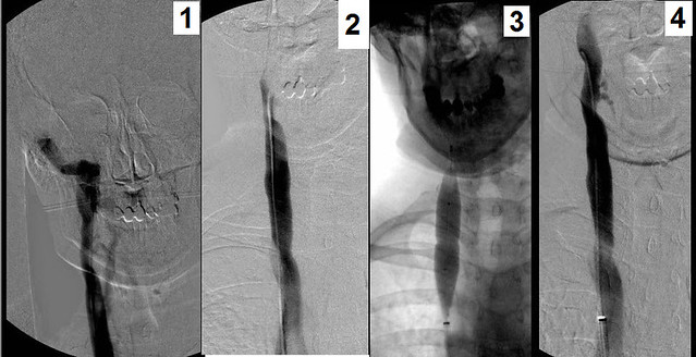
The right internal jugular vein was imaged first. The findings included clear valvular stenosis of the confluens of the IJV with the subclavian vein Figure 1, yellow circle. Simply treated by angioplasty (2), venography showed resolution of the stenosis (3)

Image
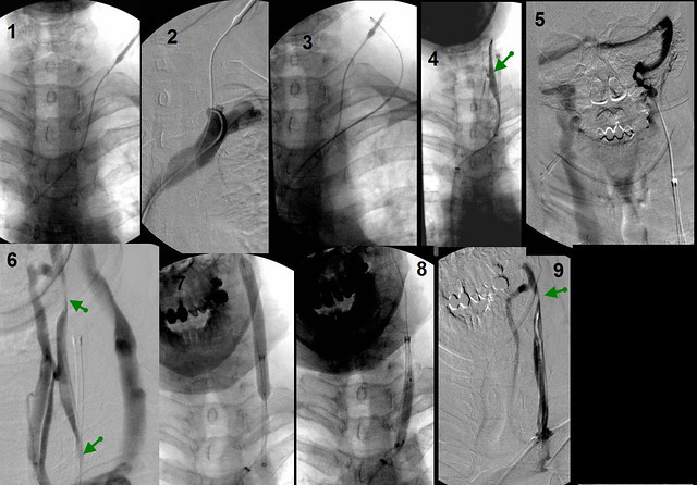
The left IJV was really prolematic and challenging. Ultrasound had shown that the vein had poor flow and was very small. As the patient had nver had angioplasty, the most likely diagnosis was hypoplasia. However recanaliation of a spontaneous occlusion was also a possibility.
After about thirty minutes of unsuccessfully trying to catheterize this vessel, i had reached my futility score.
Ultrasound of the neck was performed and I found a intermittently filled vein that was likely the IJV. So under ultrasound control, i placed a needle into the IJV. Through the needle a guidewire easilyi passed into the left inominate vein. (1). Even with the vein clearly demarkated by the jugular catheter, i could not advance the chest catheter into the jugular vein. You can see the stump of the IJV in (fig 2).

Just to make sure that the neck catheter was not in the external jugular vein, i placed the chest catheter into the external jugular vein, (3) i

The next step was to grab the chest guidewire with a snare placed through the neck sheath and pull it up above the obstruction (4). the Rendevous procedure. You can see from the figure 4, the catheter was in the IJV and some parts of it were open and others were stenotic.

That this was obstrucive cannot be denied. An injection at the skull base showed no flow going down the neck into the chest. on the contrary, all the contrast media refluxed into the dural sinuses and drained down the right jugular vein (figure 5). Pulling the catheter down and injecting contrast showed that the vein had areas of narrowing and areas of near normal size. (figure 6.)
The entire vein was angioplastied with 8 mm balloons. (fig 7-8)The final film shows a residual stenosis which remained after several dilatations. (figure 9).

So now I will place my first stent in one of my patients. it is ironic that i will have to bring my patient back for a second procedure, but when you use NO stents, you do not have much inventory on the shelf. because I use long Sheaths to introduce catheters, i need the stents are very long catheters and i just did not have one.

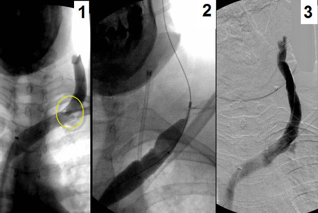
But that is not why i show this case. The conversation quoted by me at the top of this post was a question about doing angioplasty of the external jugular vein. I do not believe that there is any correlation with stenosis of the EXTERNAL jugular vein to improve hearing deficits in CCSVI. However i do believe there is a place for angioplasty but only rarely. When the patient has internal jugular obstruction, the external jugular can be an important collateral vein. When there is stenosis of the EJV, flow is impeded. Performing EJV angioplasty may improve collateral flowl.

See below
Image

Figure 1 shows stenosis of the external jugular vein (circle).
figure 2 shows balloon angioplasty
figure 3 shows no residual stenosis in the left external jugular vein

So two first on this case, a treatment of IJV occlusion by stenting and treatment of an EJV stenosis by angioplasty.
User avatar
Johnnymac
Family Elder
Posts: 213
Joined: Wed Oct 18, 2006 2:00 pm

Post by Johnnymac »

Dr S. Thanks again for everything you've done, and continue to do! Courtney is doing well, still a little sore in the chest area if she sneezes or coughs, but other than that recovery has been smooth.

A couple of things I'd like to share to date:
1) Bladder urgency seems much improved. She is no longer getting that feeling of extreme urgency, like she isn't going to make it in time. Last night she only woke once the entire evening from bladder pressure, and again it was just a 'I need to go to the bathroom' feeling, not a "I GOTTA GO NOW!" which has been the norm for a long long time.
2) Warmth and sensitivity in the her feet is improved. Her feet actually get hot now, something that used to be normal. This started the night of the procedure, and has persisted.
3) She has more sensitivity in her hands, specifically her right hand which has been unusable at time. She can feel things better, can actually cross her index and middle finger on her right hand with ease now....seems trivial but she has not been able to do that for a long time.
4) Energy. She feels much more energetic. Its hard for her to describe, but says its like a wave of energy comes over her at times, a very foreign feeling as she has been constantly fatigued for years (just getting up and dressed would wear her out).
5) Walking is pretty much unchanged at this point; however, her right leg has become much more willing to bend when she 'asks' it to. It took constant concentration and focus on her right leg when using her walker to have it bend and go where she wanted it. Now it is a little easier, the knee bends more smoothly, and with less effort.

We have a neuro visit at the end of this month, I'll definitely let you know the results of those tests and how they compare to the last one we did about 2 months ago.

If sharing images from Courtney's procedure will help anyone please feel free to do so.

Thanks again for everything!
-John & Courtney
User avatar
Needled
Family Elder
Posts: 361
Joined: Wed Jan 14, 2009 3:00 pm
Location: Connecticut
Contact:

Post by Needled »

Hi Doc,
Thought I'd go after Courtney this time so you could hear from both your patients that day, and I also wanted to thank you again for you all you do for us.
I'm doing really well, no after effects other than nasty bruises. Left foot has more feeling, back is better, and overall leg strength is better. I feel more stable and mobile. And I have an appetite. Haven't had one of those in years. So far, so good.
As John said, anything useful you want to share, please go ahead. Like maybe describing my first treatment and how it has held up. Take some credit, please!
JohnnyMac, A big hello to you and Courtney. Sounds like you're doing well. That's great to hear.
Cheryle
PS: Cece, this one was for you :wink:
Cece
Family Elder
Posts: 9335
Joined: Mon Jan 04, 2010 3:00 pm
Contact:

Post by Cece »

Thanks, Needled, I wanted to celebrate here. Fourteen months of patency! :) :)

c o n g r a t s
to doctor & patient :)
User avatar
WeWillBeatMS
Family Elder
Posts: 169
Joined: Wed Feb 03, 2010 3:00 pm
Contact:

Post by WeWillBeatMS »

Nunzio wrote:
WeWillBeatMS wrote:
drsclafani wrote:The external jugular drains the face and scalp. I do not study these veins and i do not think these veins are the first line of action, perhaps they take on a greater potential when the internal jugular cannot be opened or has clotted off.
OK. So which veins are responsible for draining the ears?
It doesn't matter; it is similar to somebody with foot drop that wants to know what vein drains the foot.
Hey Nunzio! I guess it does matter after all!

The next round of espressos are on me buddy.

How about this weekend at the Panera in CP?

WeWillBeatMS
User avatar
WeWillBeatMS
Family Elder
Posts: 169
Joined: Wed Feb 03, 2010 3:00 pm
Contact:

Post by WeWillBeatMS »

drsclafani wrote:
drsclafani wrote:
WeWillBeatMS wrote:Dr. Sclafani,

Are the external jugular veins also tested for CCSVI? I know that they have not proven to be a regular problem but what about for the people like myself who have lost hearing in one ear? And at times even my good ear rings and pops and feels plugged like my bad one does all of the time.

WeWillBeatMS
The external jugular drains the face and scalp. I do not study these veins and i do not think these veins are the first line of action, perhaps they take on a greater potential when the internal jugular cannot be opened or has clotted off.

On Saturday i treated two patients with footdrop and weakness of the lower extremity. Both had exciting reduction in foot drop within the first day. I am ever cautious to have too much excitement on early findings. the proof isin the durability of these improvements.

ONe of the patients turned into a tour de force and I would like to share with you some unusual techniques that will hopefully make a difference.

Image

The right internal jugular vein was imaged first. The findings included clear valvular stenosis of the confluens of the IJV with the subclavian vein Figure 1, yellow circle. Simply treated by angioplasty (2), venography showed resolution of the stenosis (3)


Image
The left IJV was really prolematic and challenging. Ultrasound had shown that the vein had poor flow and was very small. As the patient had nver had angioplasty, the most likely diagnosis was hypoplasia. However recanaliation of a spontaneous occlusion was also a possibility.
After about thirty minutes of unsuccessfully trying to catheterize this vessel, i had reached my futility score.
Ultrasound of the neck was performed and I found a intermittently filled vein that was likely the IJV. So under ultrasound control, i placed a needle into the IJV. Through the needle a guidewire easilyi passed into the left inominate vein. (1). Even with the vein clearly demarkated by the jugular catheter, i could not advance the chest catheter into the jugular vein. You can see the stump of the IJV in (fig 2).

Just to make sure that the neck catheter was not in the external jugular vein, i placed the chest catheter into the external jugular vein, (3) i

The next step was to grab the chest guidewire with a snare placed through the neck sheath and pull it up above the obstruction (4). the Rendevous procedure. You can see from the figure 4, the catheter was in the IJV and some parts of it were open and others were stenotic.

That this was obstrucive cannot be denied. An injection at the skull base showed no flow going down the neck into the chest. on the contrary, all the contrast media refluxed into the dural sinuses and drained down the right jugular vein (figure 5). Pulling the catheter down and injecting contrast showed that the vein had areas of narrowing and areas of near normal size. (figure 6.)
The entire vein was angioplastied with 8 mm balloons. (fig 7-8)The final film shows a residual stenosis which remained after several dilatations. (figure 9).

So now I will place my first stent in one of my patients. it is ironic that i will have to bring my patient back for a second procedure, but when you use NO stents, you do not have much inventory on the shelf. because I use long Sheaths to introduce catheters, i need the stents are very long catheters and i just did not have one.

But that is not why i show this case. The conversation quoted by me at the top of this post was a question about doing angioplasty of the external jugular vein. I do not believe that there is any correlation with stenosis of the EXTERNAL jugular vein to improve hearing deficits in CCSVI. However i do believe there is a place for angioplasty but only rarely. When the patient has internal jugular obstruction, the external jugular can be an important collateral vein. When there is stenosis of the EJV, flow is impeded. Performing EJV angioplasty may improve collateral flowl.

See below

Image

Figure 1 shows stenosis of the external jugular vein (circle).
figure 2 shows balloon angioplasty
figure 3 shows no residual stenosis in the left external jugular vein

So two first on this case, a treatment of IJV occlusion by stenting and treatment of an EJV stenosis by angioplasty.
Dr Sclafani,

Let me just go ahead and be the millionth person to remind you of how much you are changing many of our lives for the better. I haven't even had you rootin' around in my veins yet and I already feel blessed to have 'met' you here in TIMS.

Sorry I had to cancel for tomorrow and Friday. We took our little guy to the doctor again this morning and he seems to be doing better thank God.

The Mrs. and I hope to come up in July.


WeWillBeatMS
User avatar
drsclafani
Family Elder
Posts: 3182
Joined: Fri Mar 12, 2010 3:00 pm
Location: Brooklyn, New York
Contact:

Post by drsclafani »

Cece wrote:
drsclafani wrote:The entire vein was angioplastied with 8 mm balloons. (fig 7-8)The final film shows a residual stenosis which remained after several dilatations. (figure 9).

So now I will place my first stent in one of my patients.
With the vein having been hypoplastic and then if it were left with a high-up stenosis limiting blood flow, the likelihood of the vein clotting up is high. Maybe the stent prevents that outcome.
Figure 1 shows stenosis of the external jugular vein (circle).
figure 2 shows balloon angioplasty
figure 3 shows no residual stenosis in the left external jugular vein
Very nice! I'd expect external jugular vein stenoses to be along the lines of May Thurner or renal vein abnormalities, meaning no greater incidence of them in patients with MS then patients without MS but, if they are present, they may complicate the outflow in an already complicated outflow situation.

Very happy for the patients, respectively, for their foot drop improvements!!
I think that this could be a recanalized occlusion or a hypoplasia. I am betting on recanalized occlusion

Why: because the image of the confluens of the vein with the subclavian is widely patent. I would not expect that with a real hypoplasia.

Patient was never instrumented, nor had symptoms of occlusion such as pain swelling tenderness.
Cece
Family Elder
Posts: 9335
Joined: Mon Jan 04, 2010 3:00 pm
Contact:

Post by Cece »

drsclafani wrote:I think that this could be a recanalized occlusion or a hypoplasia. I am betting on recanalized occlusion.
Does a recanalized occlusion respond well to ballooning?

In this editorial, it was mentioned that a postmortem MS patient was found to have a jugular stenosis and a jugular thrombosis. Just food for thought, maybe jugular thromboses are more common in MS patients than we know. Any turbulent or stagnant blood flow could set the scene for one. It could be an explanation too for why some cases of CCSVI may worsen over time.
http://jnnp.bmj.com/content/82/4/355/reply
User avatar
Johnson
Family Elder
Posts: 979
Joined: Tue Dec 01, 2009 3:00 pm
Location: Ucluluet, BC
Contact:

Post by Johnson »

Man, I am so glad that I am going to be treated by you, Dr. Sclafani.

The idea of the contrast refluxing through the dural sinus and into the other side is freaky. It makes me think of a "nose job" with soda.

Perfect may be the enemy of good, but you are obviously way more than good. You don't give up, and that is inspiring. Great rationale for dilating the EJV. I wonder too, how many Docs would have given up when confronted with the resistance to the catheter.

Your IVUS protocol is spreading. Dr. Gilhooley at EHC is employing it now as well. (You probably know that. Just letting others know)

Karen said I couldn't have an espresso before the procedure (at 7:00 AM fer crying out loud). Could you please use espresso as a contrast medium for me? I hope you have one or two before you arrive. Laugh
My name is not really Johnson. MSed up since 1993
User avatar
magoo
Family Elder
Posts: 574
Joined: Thu Sep 10, 2009 2:00 pm
Location: Charlotte, NC
Contact:

Post by magoo »

Hi Dr. S,
I am wondering if any of your patients had pain and pressure between the shoulder blades and through to the chest after angio of the azygous? I know some soreness is normal, but I have had intense pressure and pain in these areas for 2 1/2 weeks. It does not seem to be getting better. My azygous had a thick web and a valve issue which created a 70% blockage.
In your experience, does this seem troublesome?
Thanks,
Rhonda
Rhonda~
Treated by Dake 10/19/09, McGuckin 4/25/11 and 3/9/12- blockages in both IJVs, azy, L-iliac, L-renal veins. CCSVI changed my life and disease.
User avatar
CD
Family Elder
Posts: 217
Joined: Sun Dec 19, 2010 3:00 pm
Location: USA
Contact:

Post by CD »

I moved this from Cece's turn thread..

Dr. Sclafani a question please.

I had my annual Thyroid US recently. The US women was a Dr. She found more nodules, nothing new there, and the stents in my left jugular.

She used many different US attachments to take better shots of my neck and spent a long time, one hour on me. Even going behind my ear.

She had a 3D color Doppler, but did not have me sit up or inhale, exhale. She was just surfing I think. Lol

That said, she and another Dr wrote up the report to my Endo Dr. with an addendum attached saying, I had a stent in my left jugular vein that showed "incomplete occlusion." I have tried to look this up. What does that mean? I got my copy before the Endo Dr.

I still feel the same improvements, a few came back like ringing in the ears, a few better, like balance and energy so pretty much still better, not worse.
Thank you,
CD
Where there is a will, there is a way. "HOPE"

CCSVI Procedure December 2010
newlywed4ever
Family Elder
Posts: 255
Joined: Thu Apr 17, 2008 2:00 pm
Location: Michigan
Contact:

Post by newlywed4ever »

Dr S - your last 'lesson' re: rendezvous is very exciting to me personally - we're going to get my occluded left IJV open yet...not tomorrow or the next day, but every step in this journey helps! Thanks for sharing :D
User avatar
CD
Family Elder
Posts: 217
Joined: Sun Dec 19, 2010 3:00 pm
Location: USA
Contact:

Post by CD »

Hi Magoo,
FWIW, I had Dr Siskin and he said my azygous was very tiny. The smallest he has seen so far. While he was ballooning it, a blockage at the middle, and then he did the arch, it was the pain you described. In the middle of the chest and through to my back. Ouch!

It was there as he held the balloon fully dilated. He did it many times. I must have said something or winced because he asked how it felt a few times. I said like a gallbladder attack would feel like. Sharp pain in the chest going into the shoulder blade and the back.

I was sore for a few weeks in the chest area and left shoulder blade. I am only one person, and not who you asked. I just wanted to let you know I had that too.

It went away. It is scary because you think it is heart related, but pulse and pressure were fine, while pain was still there at home. I felt like I had bruised internal ribs.
Just my story.
CD
Where there is a will, there is a way. "HOPE"

CCSVI Procedure December 2010
User avatar
drsclafani
Family Elder
Posts: 3182
Joined: Fri Mar 12, 2010 3:00 pm
Location: Brooklyn, New York
Contact:

Post by drsclafani »

magoo wrote:Hi Dr. S,
I am wondering if any of your patients had pain and pressure between the shoulder blades and through to the chest after angio of the azygous? I know some soreness is normal, but I have had intense pressure and pain in these areas for 2 1/2 weeks. It does not seem to be getting better. My azygous had a thick web and a valve issue which created a 70% blockage.
In your experience, does this seem troublesome?
Thanks,
Rhonda
hard to tell, although one has to be concerned when pain lasts that long. How big a balloon and where was the stenosis
Post Reply
  • Similar Topics
    Replies
    Views
    Last post

Return to “Chronic Cerebrospinal Venous Insufficiency (CCSVI)”