Page 435 of 557
Re: DrSclafani answers some questions
Posted: Tue Mar 13, 2012 5:59 pm
by drsclafani
drsclafani wrote:Cece wrote:ok, ok...
In the case from the stents thread that you started, did the patient have any relief of symptoms after being treated for the renal stenosis?
actually she doesnt feel so good. not sure what the problem is, fatigue from the long trip? skipping copaxone for more than a week? a relapse? an infection picked up while traveling?
we shall see
says her energy is coming back now.
Re: DrSclafani answers some questions
Posted: Tue Mar 13, 2012 6:02 pm
by drsclafani
THEGREEKFROMTHED wrote:Many patients complain of l'hermittes upon the chin to the chest. I dont get shocks but rather the stiffness increases momentarily in my lega and also the power is decreased. Can you see any possible venous connection to this phenomenon? Or could it just be excess gabagool lodged in my neck?
greek, i suspect it is stretch upon the upper cervical cord. i dont think it is a direct effect of venous obstructions,reflux or jets
s
Re: DrSclafani answers some questions
Posted: Tue Mar 13, 2012 10:04 pm
by NZer1
drsclafani wrote:THEGREEKFROMTHED wrote:Many patients complain of l'hermittes upon the chin to the chest. I dont get shocks but rather the stiffness increases momentarily in my lega and also the power is decreased. Can you see any possible venous connection to this phenomenon? Or could it just be excess gabagool lodged in my neck?
greek, i suspect it is stretch upon the upper cervical cord. i dont think it is a direct effect of venous obstructions,reflux or jets
s
Dr is this something that is CSF related?
It seems if the vascular flow is low the CSF flow is also effected and the incidence of lesions on cervical cord are common.
Is the lesions in the cervical cord a VV related thing or a CSF related?
Regards
Nigel
Re: DrSclafani answers some questions
Posted: Wed Mar 14, 2012 7:42 am
by drsclafani
NZer1 wrote:drsclafani wrote:THEGREEKFROMTHED wrote:Many patients complain of l'hermittes upon the chin to the chest. I dont get shocks but rather the stiffness increases momentarily in my lega and also the power is decreased. Can you see any possible venous connection to this phenomenon? Or could it just be excess gabagool lodged in my neck?
greek, i suspect it is stretch upon the upper cervical cord. i dont think it is a direct effect of venous obstructions,reflux or jets
s
Dr is this something that is CSF related?
It seems if the vascular flow is low the CSF flow is also effected and the incidence of lesions on cervical cord are common.
Is the lesions in the cervical cord a VV related thing or a CSF related?
Regards
Nigel
nigel
if you accept dr schelling's hypothesis, this may be a stress related problem associated with engorgement of the venous plexuses of the spine. I dont think that this is a csf problem, although that also exists and i dont think it is a vertebral vein thing, as much as it is a vertebral plexus thing which would involve any of numerous veins, including vertebral veins, azygous veins ascending lumbar veins, and renal/hemiazygous circuits
but this is hypothetical, right? But it surely sounds more like a spinal problem that csf problem
s
Re: DrSclafani answers some questions
Posted: Sat Mar 17, 2012 3:43 am
by drsclafani
It has been a while since I had the time to show an interesting case here.
This 41 year old woman visited me recently from Ankara turkey where she had previously undergone two venograms and venoplasties.
She had multiple angioplasties that resulted in clinical improvements during the first treatment. These improvements were short lasting, so she returned for another "more aggressive treatment a few months later.. She stated that no pain medications were given and that the procedure was quite painful. This treatment did not result in any return of improvements. She believed that her walking actually deteriorated after this treatment.
On my assessment she was a well dressed, organized, oriented, well speaking woman. She had weakness in both lower extremities, abnormal Rhomberg test, absent gag reflex, nystagmus and positive Babinski. Her gait was abnormal and she walked with a cane.
I performed venography which i begin to show you now.
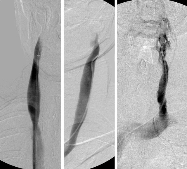
The image below shows both internal jugular veins and a vertebral vein.
Can you tell me which is which?
what abnormalities do you see??
Re: DrSclafani answers some questions
Posted: Sat Mar 17, 2012 5:14 am
by Cece
drsclafani wrote:
Oh my. The middle one is the vertebral vein but I took it for an IJV before reading your post more closely.
This is an example of what was previously believed, which was that flow would get out through one vein or another and the body would compensate. This vertebral vein is beautiful, it's as good a compensation as the body can do, but the question is if this compensation is enough, and if CCSVI symptoms are present, then it is not enough.
The right jugular in the first image looks distended in the middle and narrowed at the area of the valve. Was the flow slow? It looks like stasis. The left jugular appears blocked. Irregular appearance down at the area of the valves with dark contrast, and the usual CCSVI appearance of a cluster of small collaterals at the top of the jugular. Of the three veins, the flow looks best by far in the middle image.
A question would be if there was any obstruction in the sinus on that left side?
Do you think the patient was ballooned in the vertebral vein instead of the jugular? Are there any images from that procedure? If the patient was ballooned in the vertebral vein, did it increase the size of the vertebral vein? Where does this vertebral vein drain into? If it is draining into the vertebral plexus, then it is adding to the flow there and to the azygous vein? Was there azygous disease?
Re: DrSclafani answers some questions
Posted: Sat Mar 17, 2012 11:36 am
by Cece
Is the weather too nice and it is St Patrick's Day and no one is here to comment on the case?
drsclafani wrote:This 41 year old woman visited me recently from Ankara turkey where she had previously undergone two venograms and venoplasties.
She had multiple angioplasties that resulted in clinical improvements during the first treatment. These improvements were short lasting, so she returned for another "more aggressive treatment a few months later..
When I met with Dr. Cumming in September for a follow-up after my procedure with you in July, he was surprised (and glad) to hear that my second procedure had been more gentle than the first. He says that a common pattern among patients is to want to go more aggressive on the second procedure.
Re: DrSclafani answers some questions
Posted: Sat Mar 17, 2012 1:33 pm
by HappyPoet
Dr. S,
1) Were her pain during and lack of improvement after the second procedure indications that caused you to investigate the VVs with venography?
2) Is VV venography something you routinely/only perform when indicated?
3) Did you also perform venography on the R-VV? Average size?
Cece, great analysis, great questions!
Re: DrSclafani answers some questions
Posted: Sat Mar 17, 2012 7:33 pm
by drsclafani
Cece wrote:Is the weather too nice and it is St Patrick's Day and no one is here to comment on the case?
drsclafani wrote:This 41 year old woman visited me recently from Ankara turkey where she had previously undergone two venograms and venoplasties.
She had multiple angioplasties that resulted in clinical improvements during the first treatment. These improvements were short lasting, so she returned for another "more aggressive treatment a few months later..
When I met with Dr. Cumming in September for a follow-up after my procedure with you in July, he was surprised (and glad) to hear that my second procedure had been more gentle than the first. He says that a common pattern among patients is to want to go more aggressive on the second procedure.
Cece, i am not sure what you mean by gentler. if you mean smaller balloons or less pressure, i do not believe in that. I believe that the right size balloon at sufficient pressure to open a stenosis is appropriate. Sometimes that means increasing balloon size or increasing pressure.
Remember that the goal of second treatment is often to correct incomplete angioplasty done the first time. Of course it could also mean correcting an overly aggressive first treatment. In your case, i think would say it was really a staged treatment for a larger balloon on the first treatment would have been excessive. I have begun to believe that two treatments at planned intervals may well be a better option, rather than treatment of a re-stenosis.
It is one of the reasons that i have argued for and finally convinced Fresenius, that a global period needs to be applied to treatments. So, we will now perform planned second procedures and necessary repeats within 90 days as part of the initial fee without additional cost except for any stents that are needed. Allows my patient and I to focus on what needs to be done without worrying about unplanned costs.
I think that makes sense and is compassionate.
Re: DrSclafani answers some questions
Posted: Sat Mar 17, 2012 7:36 pm
by drsclafani
HappyPoet wrote:Dr. S,
1) Were her pain during and lack of improvement after the second procedure indications that caused you to investigate the VVs with venography?
2) Is VV venography something you routinely/only perform when indicated?
3) Did you also perform venography on the R-VV? Average size?
Cece, great analysis, great questions!
It is going to be a very educational case with the truth be quite surprising.
would you two please try to get some others to discuss this case. I would like to wait for five commentaries before discussing the answers. We all know who you are. sometime it gets lonely here.
S
Re: DrSclafani answers some questions
Posted: Sat Mar 17, 2012 11:32 pm
by NHE
The vein in the left image, possibly the right internal jugular, appears to have a twist in it that is impeding flow. The vein in the right image appears hightly disorganized at the top with contrast backfilling some collaterals. It also appears to have a potential blockage or constriction in this region. There appears to be a partial obstruction near the lower part of the vein in the image. My guess is that this could be a blocked valve or some other obstruction where it joins with another vein. Is this the left internal jugular? Was the prior treatment in the upper portion of the vein? It appears as though it may be damaged.
NHE
Re: DrSclafani answers some questions
Posted: Sun Mar 18, 2012 12:28 am
by pelopidas
drsclafani wrote:It has been a while since I had the time to show an interesting case here.
This 41 year old woman visited me recently from Ankara turkey where she had previously undergone two venograms and venoplasties.
She had multiple angioplasties that resulted in clinical improvements during the first treatment. These improvements were short lasting, so she returned for another "more aggressive treatment a few months later.. She stated that no pain medications were given and that the procedure was quite painful. This treatment did not result in any return of improvements. She believed that her walking actually deteriorated after this treatment.
On my assessment she was a well dressed, organized, oriented, well speaking woman. She had weakness in both lower extremities, abnormal Rhomberg test, absent gag reflex, nystagmus and positive Babinski. Her gait was abnormal and she walked with a cane.
I performed venography which i begin to show you now.
The image below shows both internal jugular veins and a vertebral vein.
Can you tell me which is which?
what abnormalities do you see??
normally i would vote for Cece, but i think that the vertebral vein is the third one. Is it possible, and a catheter in it?..
The patient had no real ccsvi symptoms
Plus she was well dressed !

Re: DrSclafani answers some questions
Posted: Sun Mar 18, 2012 1:02 am
by Robnl
Ok, my knowledge is not very good, but my two cents;
- I also think that the right one is the vertebral. It seems to be very 'messed up' at the top, result of aggressive treatment?
- In the lower part of the right picture there seems to be a blockage, blocked valve??
- the middle one seems ok, but maybe a twist in the lower part.
- Left one is twisted, you can see the blood 'waiting' above the twist
Dont shoot me

Robert
Re: DrSclafani answers some questions
Posted: Sun Mar 18, 2012 5:35 am
by Cece
It is a surprising case? Hmm.
Pelopidas, interesting, no real CCSVI symptoms...at least not the 'head' symptoms such as cogfog and disorientation. Weakness could be considered a CCSVI symptom. I suppose the vertebral vein could be doing a good enough job that the brain is drained, thus none of the 'brain' CCSVI symptoms, but then overloading the azygous and vertebral plexus systems, with injury to the spinal cord.
Did the patient have spinal MS lesions or brain lesions or both?
The left and right images appear to show veins that go straight up and down, like a jugular, and the middle image shows a vein that angles in toward the spinal cord, like a vertebral vein. In the right image you can also see the 'puff' of contrast into the subclavian, which looks like the jugular/innominate junction. A check of the continuity of the jugular to the dural sinus will answer the question!
So, we will now perform planned second procedures and necessary repeats within 90 days as part of the initial fee without additional cost except for any stents that are needed. Allows my patient and I to focus on what needs to be done without worrying about unplanned costs
I don't think there's another ccsvi IR out there with a policy like this. There's one that offers half-price on second treatments. This is extremely patient-friendly, and it allows you to do the treatment as you think best. I will be interested in hearing how this affects your treatments going forward. Are there particular presentations of CCSVI that might benefit most from staged treatments? Would a patient who had a less than ideal looking endpoint be brought back for a second staged treatment? This also covers anyone who has a complication from the procedure, such as clotting, or anyone whose veins close up again soon after the procedure? This will also give you more information on the results of your techniques, if you get to see previously treated patients again within 90 days.
Re: DrSclafani answers some questions
Posted: Sun Mar 18, 2012 7:19 am
by drsclafani
Cece wrote:drsclafani wrote:
Oh my. The middle one is the vertebral vein but I took it for an IJV before reading your post more closely.
This is an example of what was previously believed, which was that flow would get out through one vein or another and the body would compensate. This vertebral vein is beautiful, it's as good a compensation as the body can do, but the question is if this compensation is enough, and if CCSVI symptoms are present, then it is not enough.
The right jugular in the first image looks distended in the middle and narrowed at the area of the valve. Was the flow slow? It looks like stasis. Of the three veins, the flow looks best by far in the middle image.
A question would be if there was any obstruction in the sinus on that left side?
that "beautiful vein" in the middle is actually the left IJV. Yet it is the beast as you will see. It had excellent flow, but that flow was not from the brain
The left jugular appears blocked. Irregular appearance down at the area of the valves with dark contrast, and the usual CCSVI appearance of a cluster of small collaterals at the top of the jugular.
yes indeed but you got to the answer because you misinterpreted the enlarged vertebral vein for an internal jugular vein. The difference between good and lucky is that the person who is good knows when she is lucky. were you good or lucky, cece?
Do you think the patient was ballooned in the vertebral vein instead of the jugular? Are there any images from that procedure? If the patient was ballooned in the vertebral vein, did it increase the size of the vertebral vein? Where does this vertebral vein drain into? If it is draining into the vertebral plexus, then it is adding to the flow there and to the azygous vein? Was there azygous disease?
This very enlarged vertebral vein (one on the right) was viewed on the Ankara studies, but it was not treated. I will get into why i imaged this vein and what i did subsequently. The azygous vein was indeed diseased.