Double-checked on Haskal and here is a patient saying Haskal used IVUS on him: www.thisisms.com/ftopicp-155498.html#155498
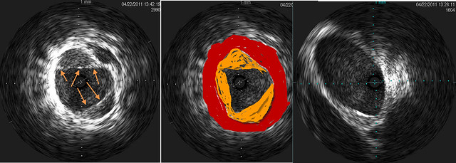
Dr. Sclafani's novel use of it to measure the stenosis to match the balloon size is unique to him, as far as I know, but I am encouraged that these other doctors have seen value in IVUS as well.
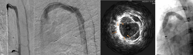
I wonder if we might get to see venogram vs ivus comparison images of any of the venograms last week (3 out of 9!) in which a stenosis was not seen on venogram but caught on IVUS? I was surprised at the mention that they were valvular stenoses. Those are big and not likely to be missed on the venogram, I would have thought.