DrSclafani answers some questions

A forum to discuss Chronic Cerebrospinal Venous Insufficiency and its relationship to Multiple Sclerosis.
User avatar
CCSVIhusband
Family Elder
Posts: 475
Joined: Sun Jun 27, 2010 2:00 pm
Location: Pittsburgh, PA USA
Contact:

Post by CCSVIhusband »

drsclafani wrote:
WeWillBeatMS wrote:Dr. Sclafani,

Are the external jugular veins also tested for CCSVI? I know that they have not proven to be a regular problem but what about for the people like myself who have lost hearing in one ear? And at times even my good ear rings and pops and feels plugged like my bad one does all of the time.

WeWillBeatMS
The external jugular drains the face and scalp. I do not study these veins and i do not think these veins are the first line of action, perhaps they take on a greater potential when the internal jugular cannot be opened or has clotted off.
Sorry to bring this topic up again ... but a few questions:

Do the IJVs drain at all when upright? Or are they completely closed off?

Do the External JVs close off too when upright?

Just curious ...
Cece
Family Elder
Posts: 9335
Joined: Mon Jan 04, 2010 3:00 pm
Contact:

Post by Cece »

CCSVIhusband, have you see this image from Dr. Zamboni? I love it for visualizing the difference between supine and upright:
www.thisisms.com/ftopicp-165261.html#165261
User avatar
CCSVIhusband
Family Elder
Posts: 475
Joined: Sun Jun 27, 2010 2:00 pm
Location: Pittsburgh, PA USA
Contact:

Post by CCSVIhusband »

I've been away for a while ... so no I hadn't. Thanks. It's very interesting ...

Still would be interested in knowing the answer to my question though ... (not sure why, just interested I guess).
User avatar
drsclafani
Family Elder
Posts: 3182
Joined: Fri Mar 12, 2010 3:00 pm
Location: Brooklyn, New York
Contact:

Post by drsclafani »

Cece wrote:
drsclafani wrote:Recently, i decided to begin my interrogation of the jugular veins by evaluating the juction between the sigmoid sinus and the jugular bulb and the jugular vein.
Does it change any of your choices during the procedure, if an issue is found there?
yes. i am in the process of writing about an experience this past weekend. Thought i had it finished last night but a glich pushed it back to today.
detecting something at the junction of jugular vein, jugular bulb and sigmoid sinus is important.
I think you just must see the entire vein.

So here it is:

I have learned many things in the past few months.
Among changes in my techniques are:
1. Venous access through the left saphenous vein using ultrasound guidance.
2. Begin the treatment by sigmoid sinus venography
3. Intravascular ultrasound
4. high pressure balloon angioplasty to a size between 50-100% above the nominal cross sectional area of the measured vein.
5. Avoidance of the use of stents except under the following theoretical circumstances:
a. restenosis after angioplasty
b. resistant stenosis
c. occlusion not responding to angioplasty
d. in-stent stenosis
e. unfavorable intimal flap or dissection

This weekend I treated a 41 year old Swiss woman with SPMS that tested a couple of these concepts.

Her initial symptoms in 1996 were optic neuritis and she went on to develop left lower extremity, left upper extremity and truncal weakness associated with spasticity. She lacks balance, has severe fatigue and heat intolerance, and suffers from migraine-like headaches. She has urinary urgency and constipation. After 10 years of copaxone, she currently is on no disease modifying medications.

In March 2010 she underwent her first CCSVI treatment in an eastern European country. At that time, stenosis of the lower right internal jugular vein was treated by a 10 mm angioplasty followed by 12 mm stenting. She received no benefit from this procedure.
Image
right IJVenography showed a 50% stenosis of the right IJV at confluens with residual stenosis after 10 mm angioplasty. Thus a stent was place (12mm by 4cm) in the J1 segment. The stent was low and covered the origin of the right subclavian vein.
Note that the upper J3 segment of the vein appears assymetrically narrower than the remainder of the jugular vein. No treatment was offered.


Image
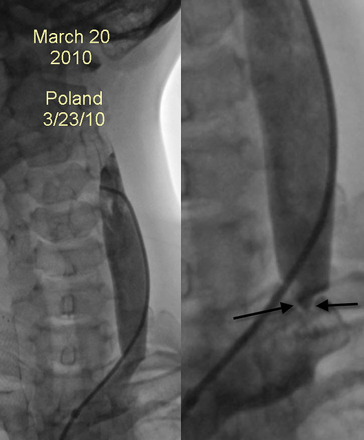
The image on the left looks unremarkable but on closer review of other images in the sequence (IMAGE ON RIGHT), a jet, suspicious for a high grade stenosis of the confluens is seen. Unfortunately, it was not recognized at that time and no angioplasty was performed. Moreover the left upper jugular vein was not visualized.


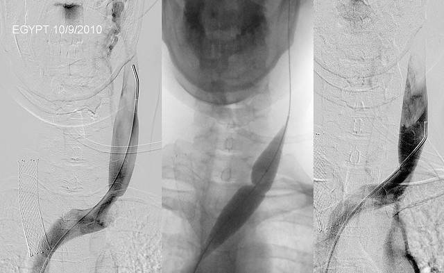
In November 2010 she had her procedure repeated in Alexandria .

Image
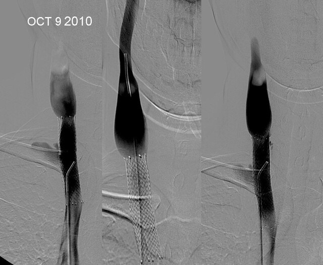
The examination in Egypt showed some intimal hyperplasia of the junction of the stent and the nonstented vein above the stent. The upper IJV was not imaged. After angioplasty, the mild intimal hyperplastic narrowing was no longer seen. No additional stenting was performed.

Image
The high grade stenosis underestimated in march 2010 was again seen. Her left internal jugular vein was treated by 12 mm angioplasty with a cutting wire ("poor man's cutting balloon"). It appears that this angioplasty was considered satisfactory.

Image
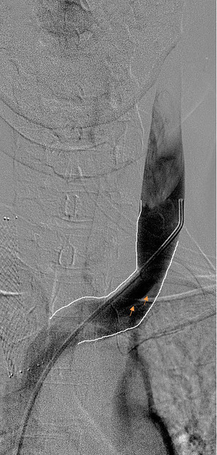
However venography clearly shows persistent stenosis (ARROWS).


Image
Angioplasty of the azygous vein was also performed.

She received limited and short term relief.


It is not my intention to show others' errors. We ALL made mistakes and misinterpretations and probably continue to do so as this is a different presentation that we all assumed at the beginning. I remember thinking that this was simple, and looking at dr Zamboni's papers i felt justified in thinking this. But with time, the subtle nature of some of the abnormal imaging presentations have become clearer.

So having set the table, I share with you my experience.

As you know, I still find it difficult to complete three cases in a day, but feel compelled to treat as many patients as possible, safely and accurately. So it was with some satisfaction that i had completed my second procedure on Friday by 1pm with the aim of leaving the center by the decent time of 5pm. Would that it were true!

I initially catheterized the right internal jugular vein.

Image
there is mild intimal hyperplasia at the top of the stent resulting in minor stenosis. However note that the upper reaches of the right IJV are not visualized. Attempts to advance a guidewire only resulted in mild contrast extravasation. There was very good flow but it was unclear whether this was from collateral vessels entering the IJV or from direct flow from the head.

i left the right jugular with an intention to return there depending upon what i found in the other vessels.

Left IJV venography was next performed.

Image
Initially the guidewire was advanced through the left jugular bulb into the sigmoid sinus. Then it was advanced across the transverse sinus, into the right sigmoid sinus and finally past the right jugular bulb and into the the jugular vein.

I finally knew the location of the highest part of the right internal jugular vein and that there was a lumen of the vein.

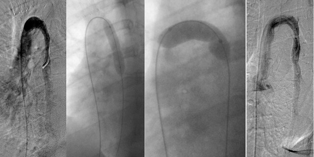
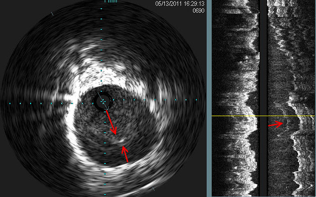
before returning to the RIJV, left IJV venography was performed. It showed a subtle "jet" of contrast media (red arrows). Intravascular ultrasound showed a septum across the ostium of the left IJV at its entrance into the left subclavian vein. on the sagittal view, the septum was represented as scattered white lines (red arrow) on IVUS

Venoplasty clearly shows the "waist" on the balloon and after 14 atmosphered of pressure were applied, the "waist was no longer seen. I wonder whether this will also represent a challenge as Nunzio's left jugular was!

Image

Now i returned to the right IJV. repeated attempts to advance the guidewire were performed for several minutes with no success. Then the guidewire advanced with a push up and into the jugular bulb and canal. Eventually the wire was placed within the right sigmoid sinus and IVUS was done.

Image
Ivus showed that there was occlusion of the upper segment of the RIJV.

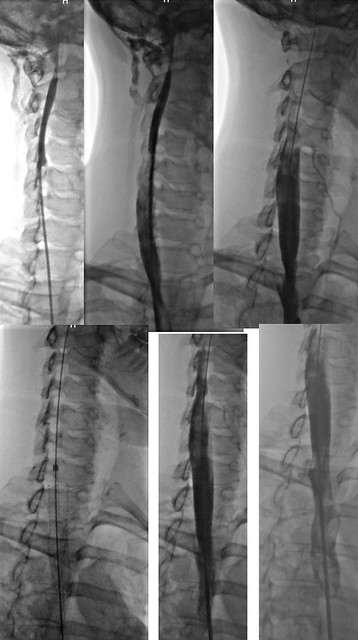
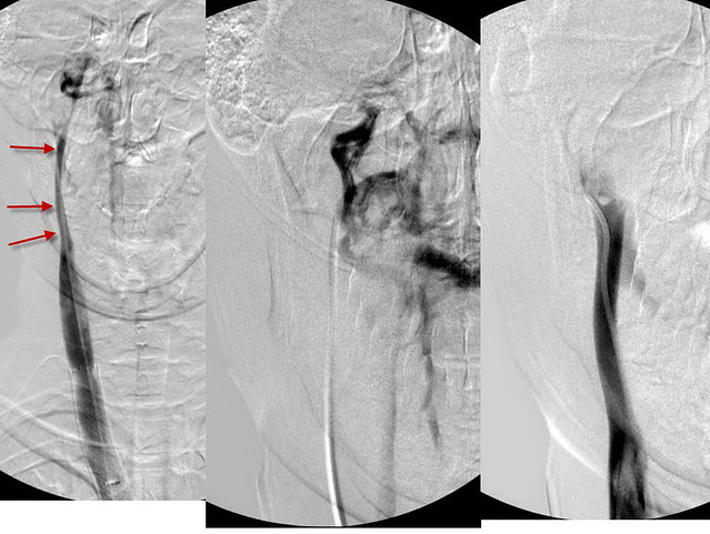
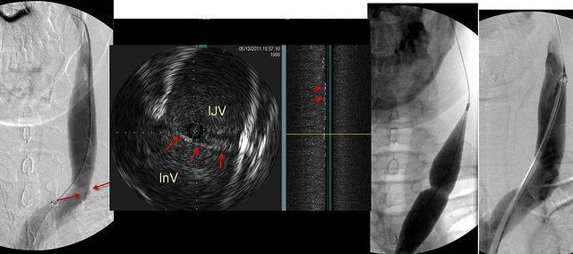
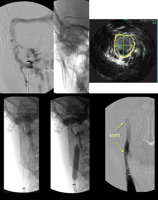
The next image is a composite that summarizes the intervention done on the J3 segment of the RIJV

Image
UPPER LEFT image shows contrast in the sigmoid sinus. However the upper jugular vein was not seen. Collaterals in the neck and spine were seen.
UPPER MID: A second wire was advanced as a safety wire and over it was placed an IVUS probe. (dark spot)
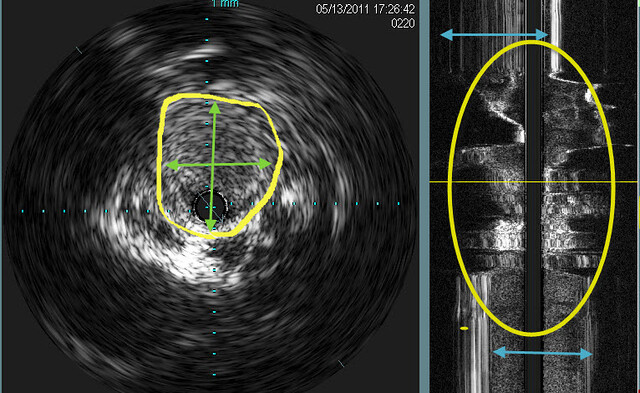
UPPER RIGHT: IVUS shows an occlusion of the upper jugular vein. Measurment of diameters of occlusions is not easy by venography, but IVUS is quite precise (YELLOW ARROWS).
LOWER LEFT & MIDDLE: 8mm by 6cm angioplasty performed. However there was no flow on angiography.
LOWER RIGHT: Therefore a stent was placed8mm by 4cm. Venography shows flow.

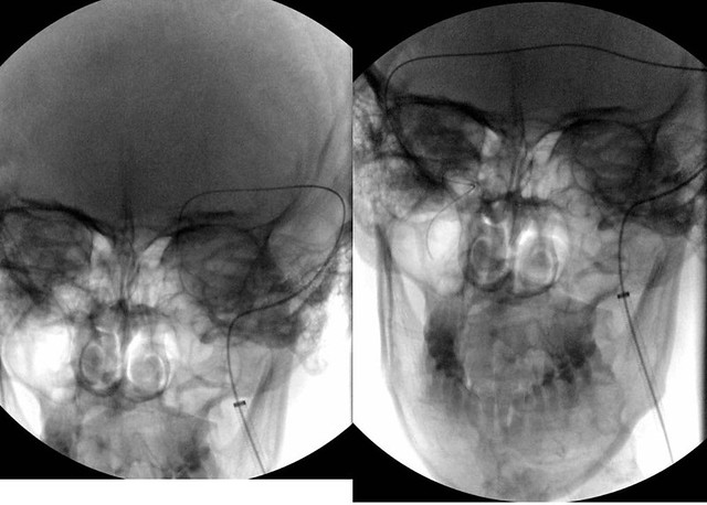
Finally, we imaged the azygous vein.

Image

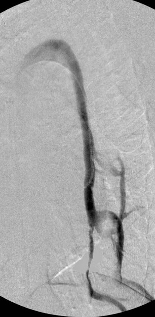
the image looked pretty good to me. However there was a persistent immobile valve that was treated by balloon angioplasty.

ImageFinally in the ascending azygous vein i noted some funky looking echoes. These likely represent webs.

So that the story of my patient and me on a late Friday night date.

hopefully she will receive some benefits of this.

Things i learned are that
1. prior treatments may not be durable
2. Early treatments overlook things
3. the entire jugular vein must be imaged
4. there is a role for stents
5. webs may be visible on IVUS

any questions?
Last edited by drsclafani on Mon May 16, 2011 4:13 pm, edited 1 time in total.
User avatar
pklittle
Family Elder
Posts: 341
Joined: Sun Mar 07, 2010 3:00 pm

Post by pklittle »

Dr., will you please let us know if/when this lady reports benefits? I am particularly interested in knowing about this case. I assume she does not post here(?)

It is great to read about all the new things you are discovering!
User avatar
Thekla
Family Elder
Posts: 149
Joined: Sun Jul 04, 2010 2:00 pm
Location: Germany

Post by Thekla »

I would love to know how the results for her are. It seems that we are hearing more often of stent problems from stents that maybe ought not to have been placed and even more often of problems overlooked or unrecognized by the early treating doctors. Do you think that doctors are, across the board, getting the experience and adapting their procedures or is this a future conference lecture.

You mention a 'persistent immobile valve' in the azygous that you treated by angioplasty. Was that just not identified in her previous procedures?

Are you finding the sigmoid sinus cropping up more now? Somehow, the idea of a wire there really creeps me out.

It seems like there is so much to learn with this. I really hate having to be at the beginning of the learning process---I wonder what we'll know in another year or two.
Cece
Family Elder
Posts: 9335
Joined: Mon Jan 04, 2010 3:00 pm
Contact:

Post by Cece »

Among changes in my techniques are:
1. Venous access through the left saphenous vein using ultrasound guidance.
This is to avoid complications with the femoral vein, such as the femoral thrombosis that another IR's patient experienced. It's very uncommon, isn't it, to enter through the saphenous vein, for any procedure? Is it part of the rationale that many CCSVI patients are having multiple procedures and therefore multiple opportunities to damage the femoral vein? If the saphenous vein is damaged, it is not as serious.
5. Avoidance of the use of stents except under the following theoretical circumstances:
a. restenosis after angioplasty
b. resistant stenosis
c. occlusion not responding to angioplasty
d. in-stent stenosis
e. unfavorable intimal flap or dissection
In these cases, the risk of stents is acceptable? I am not convinced....
With travelling patients, they may not get any follow-up care for those stents.
I have no argument with (d.), use of a stent to treat in-stent stenosis!
User avatar
phe
Getting to Know You...
Posts: 14
Joined: Fri Oct 22, 2010 2:00 pm

Post by phe »

Yikes...the case above is the one you had after I kneed you for 3 hrs Friday? And I thought I was challenging.

Here's a wee benefit with mixed emotions...my spasticity is gone for now...yay...but I can't use it to stand up...so rehab it is.

Dr S...you are my hero!
User avatar
drsclafani
Family Elder
Posts: 3182
Joined: Fri Mar 12, 2010 3:00 pm
Location: Brooklyn, New York
Contact:

Post by drsclafani »

phe wrote:Yikes...the case above is the one you had after I kneed you for 3 hrs Friday? And I thought I was challenging.

Here's a wee benefit with mixed emotions...my spasticity is gone for now...yay...but I can't use it to stand up...so rehab it is.

Dr S...you are my hero!
This is incredible. I would have bet all the equity in my house that you would not have had any reduction is your spasticity. It was so bad i had to do the procedure holding your flexed leg with my body while i worked around your leg.

i am so happy tonite!

DrS
User avatar
drsclafani
Family Elder
Posts: 3182
Joined: Fri Mar 12, 2010 3:00 pm
Location: Brooklyn, New York
Contact:

Post by drsclafani »

Cece wrote:
Among changes in my techniques are:
1. Venous access through the left saphenous vein using ultrasound guidance.
This is to avoid complications with the femoral vein, such as the femoral thrombosis that another IR's patient experienced. It's very uncommon, isn't it, to enter through the saphenous vein, for any procedure? Is it part of the rationale that many CCSVI patients are having multiple procedures and therefore multiple opportunities to damage the femoral vein? If the saphenous vein is damaged, it is not as serious.
5. Avoidance of the use of stents except under the following theoretical circumstances:
a. restenosis after angioplasty
b. resistant stenosis
c. occlusion not responding to angioplasty
d. in-stent stenosis
e. unfavorable intimal flap or dissection
In these cases, the risk of stents is acceptable? I am not convinced....
With travelling patients, they may not get any follow-up care for those stents.
I have no argument with (d.), use of a stent to treat in-stent stenosis!
Cece
are you saying that a stenosis or occlusion is better than a stent? All of these problems result in little to no flow through the jugular vein.

They are also very rarely encountered.

with respect to long distance travelers, it is possible to have patients with stents cared for safely as long as you partner with the patient's primary physician (or at least another IR.) :wink:
User avatar
drsclafani
Family Elder
Posts: 3182
Joined: Fri Mar 12, 2010 3:00 pm
Location: Brooklyn, New York
Contact:

Post by drsclafani »

pklittle wrote:Dr., will you please let us know if/when this lady reports benefits? I am particularly interested in knowing about this case. I assume she does not post here(?)

It is great to read about all the new things you are discovering!
she occasionally posts here. It is up to her if she wants to post, obviously.

i will share any information i receive. As we know, all the IR can do is treat to best ability. Outcomes then become up to the patient and the disease.
Cece
Family Elder
Posts: 9335
Joined: Mon Jan 04, 2010 3:00 pm
Contact:

Post by Cece »

drsclafani wrote:Cece
are you saying that a stenosis or occlusion is better than a stent? All of these problems result in little to no flow through the jugular vein.

They are also very rarely encountered.

with respect to long distance travelers, it is possible to have patients with stents cared for safely as long as you partner with the patient's primary physician (or at least another IR.) :wink:
It is possible but is it plausible? Canada....
But I am glad to hear they are rarely encountered.

It may be that a stenosis or occlusion may become treatable as advancements are made; this is preferable to a stent that may become untreatable (due to clotting, occlusion, the usual), to me, having read a lot of bad outcomes with stents and being early, myself, in the disease process.

I have only gotten so far as responding to the first two paragraphs of your post! Glad to see you enlargened the pictures. My vision improved since venoplasty, but not quite that much. :)
User avatar
drsclafani
Family Elder
Posts: 3182
Joined: Fri Mar 12, 2010 3:00 pm
Location: Brooklyn, New York
Contact:

Post by drsclafani »

dania wrote:Dr Sclafani I have a stent that was inserted last June. It has now become pinched and bent at one end. Dr Arata was unable to get past the bend. It is causing me considerable pain. Meds do not help. What can be done?Can it be removed?
i would need to see the pictures before rendering an opinion
User avatar
drsclafani
Family Elder
Posts: 3182
Joined: Fri Mar 12, 2010 3:00 pm
Location: Brooklyn, New York
Contact:

Post by drsclafani »

CCSVIhusband wrote:
drsclafani wrote:
WeWillBeatMS wrote:Dr. Sclafani,

Are the external jugular veins also tested for CCSVI? I know that they have not proven to be a regular problem but what about for the people like myself who have lost hearing in one ear? And at times even my good ear rings and pops and feels plugged like my bad one does all of the time.

WeWillBeatMS
The external jugular drains the face and scalp. I do not study these veins and i do not think these veins are the first line of action, perhaps they take on a greater potential when the internal jugular cannot be opened or has clotted off.
Sorry to bring this topic up again ... but a few questions:

Do the IJVs drain at all when upright? Or are they completely closed off?

Do the External JVs close off too when upright?

Just curious ...
in most cases there is always some flow in the jugular vein
i do not think that the EJV are positional in their flow
User avatar
drsclafani
Family Elder
Posts: 3182
Joined: Fri Mar 12, 2010 3:00 pm
Location: Brooklyn, New York
Contact:

Post by drsclafani »

Cece wrote:
Among changes in my techniques are:
1. Venous access through the left saphenous vein using ultrasound guidance.
This is to avoid complications with the femoral vein, such as the femoral thrombosis that another IR's patient experienced. It's very uncommon, isn't it, to enter through the saphenous vein, for any procedure? Is it part of the rationale that many CCSVI patients are having multiple procedures and therefore multiple opportunities to damage the femoral vein? If the saphenous vein is damaged, it is not as serious.
the reasons are several
1. injury to the femoral vein has greater consequences
2. repeated punctures might lead to stricture.
3. always do the most bengn and less risky procedure.
4. the femoral vein is closer to the femoral artery
Post Reply
  • Similar Topics
    Replies
    Views
    Last post

Return to “Chronic Cerebrospinal Venous Insufficiency (CCSVI)”