DrSclafani answers some questions

A forum to discuss Chronic Cerebrospinal Venous Insufficiency and its relationship to Multiple Sclerosis.
Cece
Family Elder
Posts: 9335
Joined: Mon Jan 04, 2010 3:00 pm
Contact:

Re: DrSclafani answers some questions

Post by Cece »

drsclafani wrote:Image
There's picture #8 to which HappyPoet is referring. :)
I can't tell what is going on in picture #5.
Pictures #6 & 7 would be the predilating? There are waists evident that did not resolve, but I don't suppose that matters what a stent is going in.
User avatar
NHE
Volunteer Moderator
Posts: 6238
Joined: Sat Nov 20, 2004 3:00 pm
Contact:

Re: DrSclafani answers some questions

Post by NHE »

HappyPoet wrote:Regarding picture No. 8, what does the indentation on the left side in the upper stent signify and what factors might account for that portion of the stent not fully opening?
Cece wrote: There are waists evident that did not resolve, but I don't suppose that matters what a stent is going in.
The indentations in the stent appear to correspond to the locations of the waists. Although the magnification is different, the waists appear to be located next to the same bones as the indentations in the stent.


NHE
User avatar
mo_en
Family Member
Posts: 48
Joined: Thu Dec 09, 2010 3:00 pm
Location: Patras, Greece

Re: DrSclafani answers some questions

Post by mo_en »

How different is the anticoagulation regime after that kind of stenting?

And something else. A patient that has already undergone 2 procedures the last three months (June and September) with no improvement but with serious deterioration of both CCSVI and MS symptoms, how long should wait before a new attempt (wishfully with you this time)?
Cece
Family Elder
Posts: 9335
Joined: Mon Jan 04, 2010 3:00 pm
Contact:

Re: DrSclafani answers some questions

Post by Cece »

mo_en wrote:A patient that has already undergone 2 procedures the last three months (June and September) with no improvement but with serious deterioration of both CCSVI and MS symptoms, how long should wait before a new attempt (wishfully with you this time)?
Serious deterioration? Is it known whether there are complications such as clotting, or if this is deterioration despite good flow?
User avatar
drsclafani
Family Elder
Posts: 3182
Joined: Fri Mar 12, 2010 3:00 pm
Location: Brooklyn, New York
Contact:

Re: DrSclafani answers some questions

Post by drsclafani »

CD wrote:Dr S. I am trying to find out the lifespan of stents. I have 3 Ev3 mesh titanium with nickle alloy stents placed in my left IJV. They do overlap each other from two procedures. Mine look similar to your above image with the stent/s. Mine may go even higher. I have a small neck. You can see and feel them. :?

Cece said
I don't think we know yet what the lifespan of stents will be when placed in the jugulars. They may be subject to more movement stress here than in more stable areas of the body. I can remember one person reporting that they'd had a stent fracture but only that one person
What is your opinion please?
CD
The risk of fracture of a stent in the jugular vein is impossible to state. I could find no instances in theliterature;however, there really are few stents in the jugulars to begin with. The most common fracture reported is stents within the coronary arteries, not surprising since the most stents are placed in this artery. Originally the incidence was reported to be about 2-4 % but most believe that the incidence is higher.

Leg artery stents have a much higher reported incidence. One report quoted a 37% frequency.
Other reports found a 28% fracture rate in one year
another report was a 17% four year fracture rate.

Fractures stents in arteries are associated with a 1/3 restenosis rate and a one third thrombosis rate
Salvatore JA Sclafani MD
Patient contact: ccsviliberation@gmail.com
User avatar
drsclafani
Family Elder
Posts: 3182
Joined: Fri Mar 12, 2010 3:00 pm
Location: Brooklyn, New York
Contact:

Re: DrSclafani answers some questions

Post by drsclafani »

HappyPoet wrote:DrS, it's wonderful that your rendezvous procedure was successful in helping to open the vein for stenting. Your success will give hope to those who lost their improvements when their IJVs closed.

What type of blockages are the most difficult to get through with guidewires: blood clots, intimal hyperplasia, or scar tissue?
in order of difficulty (least to greatest difficulty):
intimal hyperplasia
acute thrombus (clot)
dissection of the wall
chronic clot
scar tissue
What factors contribute to how difficult a blockage is to get through with the guidewires?
The absence of a virtual lumen makes it really difficult. There is a lumen present in soft young clot. There is a lumen in intimal hyperplasia. Dissections make it difficult especially when the flap directs the guidewire into the wall rather than into the lumen. chronic clot may have no residual lumen or the lumen is discontinuous. the wire in scar in the vein just has no place to go without poking thru the scar.
Also, is there any kind of rendezvousesque procedure available for the Azygos vein?
It' a possibility through collaterals but i really do not think it would work. it was through the left renal vein, through the innominate vein or through the ascending lumbar vein. I hope I never have to try that because there really is no likely success.
Regarding picture No. 8, what does the indentation on the left side in the upper stent signify and what factors might account for that portion of the stent not fully opening? Suddenly occurring to me is what about the other side of the stent that can't be seen? Can IRs see the outside of the vein/stent from all sides?
it means that we didnt store a picture after angioplasty of the stent to smooth out the expansion. Remember we are going through scar tissue, not a vein.
Edit: Do you have any images that show the tips of the two guidewires (one wire approaching from above the clot and the other wire approaching from below the clot) close to each other or meeting? Thanks!
somewhere, but sorry, i do not have time to search for it. I'll show it the next time i help another patient with this problem.
Salvatore JA Sclafani MD
Patient contact: ccsviliberation@gmail.com
User avatar
drsclafani
Family Elder
Posts: 3182
Joined: Fri Mar 12, 2010 3:00 pm
Location: Brooklyn, New York
Contact:

Re: DrSclafani answers some questions

Post by drsclafani »

Cece wrote:
drsclafani wrote:Image
There's picture #8 to which HappyPoet is referring. :)
I can't tell what is going on in picture #5.
happy poet! picture #5 shows the snare in the sheath pulling up the wire from below. not easy to recognize.
Pictures #6 & 7 would be the predilating? There are waists evident that did not resolve, but I don't suppose that matters what a stent is going in.
sometimes, for sake of time, i will only show the balloon showing the waist on the balloon.
Salvatore JA Sclafani MD
Patient contact: ccsviliberation@gmail.com
User avatar
drsclafani
Family Elder
Posts: 3182
Joined: Fri Mar 12, 2010 3:00 pm
Location: Brooklyn, New York
Contact:

Re: DrSclafani answers some questions

Post by drsclafani »

mo_en wrote:How different is the anticoagulation regime after that kind of stenting?
anticoagulation after stenting must continue until the stent is incorporated into the wall. Generally antiplatelet and antithrombin agents are used. Different regimens are used. Some prefer an anticoagulant and two antiplatelet agents. My preferences are aspirin and dabigatran (pradaxa), a twice daily capsule. alternative is coumadin, more difficult to manage, but less expensive.
And something else. A patient that has already undergone 2 procedures the last three months (June and September) with no improvement but with serious deterioration of both CCSVI and MS symptoms, how long should wait before a new attempt (wishfully with you this time)?
some CCSVI IRs say that if the first treatment is not successful, stop.
I think that is unfair to a patient with no options. I am willing to repeat the procedure looking with IVUS for things that have gone unnnoticed without it, or perhaps using larger balloons.

But two prior treatments? Before I would commit, i would have to review all the prior efforts in great detail, images, talk to IR, etc.

it seems that i am the backup guy! I would say that half the patients coming to see me have already had the procedure. I would prefer to take the first shot at this. but no one gets refused consideration.
Salvatore JA Sclafani MD
Patient contact: ccsviliberation@gmail.com
User avatar
drsclafani
Family Elder
Posts: 3182
Joined: Fri Mar 12, 2010 3:00 pm
Location: Brooklyn, New York
Contact:

Re: DrSclafani answers some questions

Post by drsclafani »

mo_en wrote:How different is the anticoagulation regime after that kind of stenting?

And something else. A patient that has already undergone 2 procedures the last three months (June and September) with no improvement but with serious deterioration of both CCSVI and MS symptoms, how long should wait before a new attempt (wishfully with you this time)?
sorry, didnt complete answer. If you are getting worse, i would make sure you have a recent neurological assessment and an MRI looking for more active lesions. Perhaps you are highly inflamed and need to be more patient. if lesional activity is not worsening, i might consider early treatment
Salvatore JA Sclafani MD
Patient contact: ccsviliberation@gmail.com
User avatar
drsclafani
Family Elder
Posts: 3182
Joined: Fri Mar 12, 2010 3:00 pm
Location: Brooklyn, New York
Contact:

Re: DrSclafani answers some questions

Post by drsclafani »

Cece wrote:
mo_en wrote:A patient that has already undergone 2 procedures the last three months (June and September) with no improvement but with serious deterioration of both CCSVI and MS symptoms, how long should wait before a new attempt (wishfully with you this time)?
Serious deterioration? Is it known whether there are complications such as clotting, or if this is deterioration despite good flow?
yes, of course. an ultrasound should have already been done to look for clotting. I am assuming that there is no clot, for no good reason
Salvatore JA Sclafani MD
Patient contact: ccsviliberation@gmail.com
User avatar
drsclafani
Family Elder
Posts: 3182
Joined: Fri Mar 12, 2010 3:00 pm
Location: Brooklyn, New York
Contact:

Re: DrSclafani answers some questions

Post by drsclafani »

Bluesky spoke about a possibility of pelvic congestion syndrome recently. So i wanted to review this case that was performed yesterday.

The patient is a 52 year old canadian nurse with weakness and spasticity in the lower extremities, accompanied by pain, cog fog, memory issues, heat intolerance and chronic fatigue.
She had a six vessel evaluation. I am showing all the abnormal findings on venography only because of time constraints. IVUS showed that stenoses were all due to valvular immobility and stenosis.
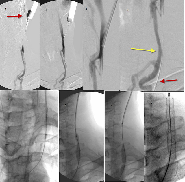
RIGHT IJV
Image
There is a valvular stenosis at the confluens with bulging of the right sided valve cusp. 18 mm high pressure balloon used. 8 atmospheres needed to open. Results looked very good to me.

LEFT IJV
Image
There is valvular stenosis at the confluens. Neither the cusps nor the valve leaflets were visible on venography but valve stenosis was seen on IVUS. Angioplasty with 14 mm high pressure balloon was used. Pressure was 15 Atmospheres

AZYGOUS VEIN
Image

there was very high flow in the azygous vein and irregularities in the azygous arch. IVUS showed two immobile valves on the arch. A 14 mm balloon was used to 6 atmospheres with good result. IVUS showed no valve problem after treatment. But flow was very strong and the vein itself was very large, suggesting to me that we were dealing with a Nutcracker syndrome.

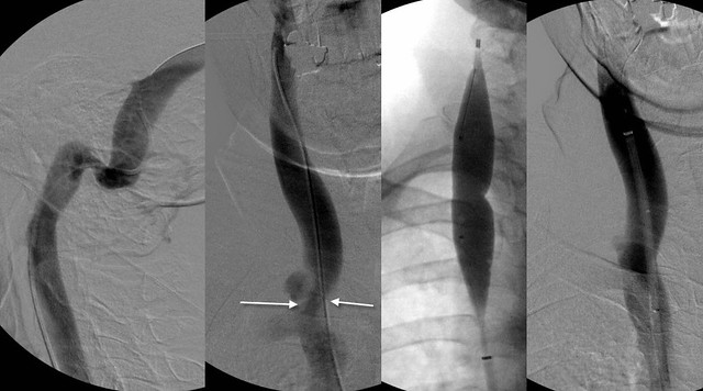
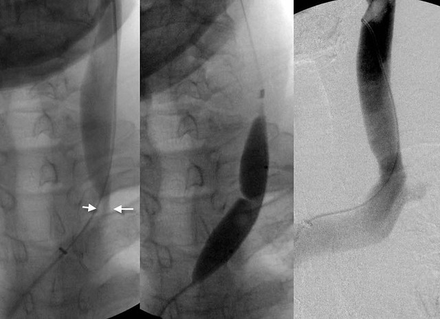
RENAL VEIN AND ASCENDING LUMBAR VEIN
Image

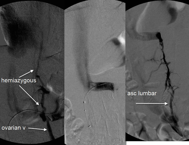
The renal venogram shows a classic Nutcracker syndrome. The contrast is "light" as the vein nears the inferior vena cava. this is caused by the compression. There is dense filling of both the ovarian vein and the hemiazygous vein which are being used as collaterals to drain the left kidney of its high blood flow volume. On the left is seen the ascending lumbar vein. this also connects to the renal vein.

RNutcracker syndrome may be a clinically occult problem in healthy patients but in patients with compromised outflow of the azygous vein, additional blood flow enters the vertebral plexus and the spinal veins. thus compounds the CCSVI resulting from the stenoses of the right and left IJV and the azygous vein.

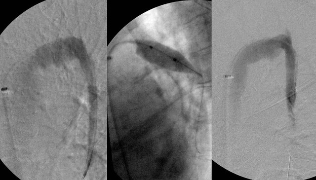
After the competion of the procedure the patient upon questioning volunteered information regarding chronic pain near the left ovary that was, to her reminiscent of pain in the ovary during menstruation. (she is post menopausal). Thus i believe that she has pelvic congestion caused by reflux of renal vein blood into her ovarian vein resulting from the nutcracker syndrome.

It will be interesting to see whether her ovarian symptoms improve now that the renal vein has been opened. However the high flow down the ovarian vein has resulted in incompetence of the valves of her ovarian vein and she may not get relief without treatment of the ovarian vein syndromed.

The treatment is blockade of the ovarian vein from the flow in the renal vein by embolization.

Hope this is helpful bluesky

DrS
Salvatore JA Sclafani MD
Patient contact: ccsviliberation@gmail.com
Cece
Family Elder
Posts: 9335
Joined: Mon Jan 04, 2010 3:00 pm
Contact:

Re: DrSclafani answers some questions

Post by Cece »

drsclafani wrote:The risk of fracture of a stent in the jugular vein is impossible to state. I could find no instances in theliterature;however, there really are few stents in the jugulars to begin with. The most common fracture reported is stents within the coronary arteries, not surprising since the most stents are placed in this artery. Originally the incidence was reported to be about 2-4 % but most believe that the incidence is higher.

Leg artery stents have a much higher reported incidence. One report quoted a 37% frequency.
Other reports found a 28% fracture rate in one year
another report was a 17% four year fracture rate.

Fractures stents in arteries are associated with a 1/3 restenosis rate and a one third thrombosis rate
NHE linked to this awhile back http://bibamed.agcl.com/cx_2005/1620Scheinert.pdf
It shows some disturbing images of stent fractures in an artery.
And on p. 26, there was a good illustration of the different forces that a stent must withstand (extension/contraction, torsion, compression, flexion).

It's possible that a stent could fracture and continue to do its job, according to this guy
http://www.thisisms.com/forum/chronic-c ... tml#p99459

But what I was looking for was a report we heard once, I believe it was from someone who had had an undersized stent placed, and their IR planned to deliberately fracture it to stretch it and then place a correctly sized stent? Does that sound familiar to anyone?
Cece
Family Elder
Posts: 9335
Joined: Mon Jan 04, 2010 3:00 pm
Contact:

Re: DrSclafani answers some questions

Post by Cece »

drsclafani wrote: in order of difficulty (least to greatest difficulty):
intimal hyperplasia
acute thrombus (clot)
dissection of the wall
chronic clot
scar tissue
This was helpful, I didn't have a clear sense of what order they'd be in.
Cece
Family Elder
Posts: 9335
Joined: Mon Jan 04, 2010 3:00 pm
Contact:

Re: DrSclafani answers some questions

Post by Cece »

drsclafani wrote: RIGHT IJV
Image
There is a valvular stenosis at the confluens with bulging of the right sided valve cusp. 18 mm high pressure balloon used. 8 atmospheres needed to open. Results looked very good to me.

LEFT IJV
Image
There is valvular stenosis at the confluens. Neither the cusps nor the valve leaflets were visible on venography but valve stenosis was seen on IVUS. Angioplasty with 14 mm high pressure balloon was used. Pressure was 15 Atmospheres
I would think an 18 mm balloon on one side and a 14 mm balloon on the other side means that she has large jugulars, overall. Were these large jugulars or about typical? Do you think there might be a better outcome in terms of symptom improvement in patients with a greater total jugular CSA after the procedure?

Dr. Tucker was saying in another thread that men have greater elasticity in their jugulars than women do. Would that mean that female jugulars are more prone to injury if overstretched by ballooning? It also seemed to mean that male jugulars, being elastic, would have a greater capacity as holding tanks which would mean less reflux. I wonder if anything can be done (or medication taken) as an adult to increase the elasticity of the jugulars.

I notice you used a 15 atm balloon on the second jugular. Is it relatively rare to need to go that high? What is the highest that you are currently comfortable going?
User avatar
Squeakycat
Family Elder
Posts: 411
Joined: Fri Dec 04, 2009 3:00 pm
Location: Yehud, Israel

Re: DrSclafani answers some questions

Post by Squeakycat »

Cece wrote:
drsclafani wrote: Dr. Tucker was saying in another thread that men have greater elasticity in their jugulars than women do. Would that mean that female jugulars are more prone to injury if overstretched by ballooning? It also seemed to mean that male jugulars, being elastic, would have a greater capacity as holding tanks which would mean less reflux. I wonder if anything can be done (or medication taken) as an adult to increase the elasticity of the jugulars.
With women more susceptible to MS than men, I wonder if differences like this in basic anatomy play a role in that differentiation?
Post Reply
  • Similar Topics
    Replies
    Views
    Last post

Return to “Chronic Cerebrospinal Venous Insufficiency (CCSVI)”