Cece wrote:
drsclafani wrote:The "messy" area looks like reflux under an obstructed valve to me.
You had described this, using a wine glass in a tumbler image, at the symposium patient day. Here was me trying to describe what you had said:
http://www.thisisms.com/forum/chronic-c ... ml#p171050
I bet there's a link out there for the actual presentation now....
so lets review the right internal jugular venograms. I will review all the IVUS studies together later:
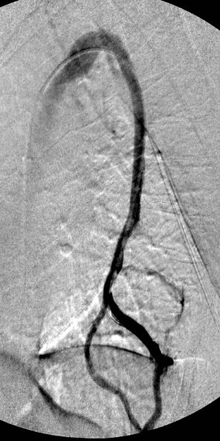
This vein was treated successfully a year ago. This image shows that there is a narrowing of the mid right IJV. Below this there is some irregularity and bulging of the contrast at the area ofof previously treated valve problems. There is reflux into the right external jugular vein (to the left of the IJV on the image).
Now a composite summary of the right side.
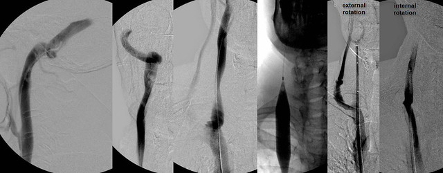
The first two images show the right transverse sinus emptying into the internal jugular vein. I think that this is the optimum way to fully evaluate the internal jugular vein. Opacify it by putting the constrast media upstream from the jugular vein. Sometimes, not often, you will see something wrong with the dural sinuses. You will also get a long at the condylar emissary vein which is a often seen collateral vein.Collaterals seen are minimal.
There was evidence of stenosis at the confluens where the valve was treated previously. There is refluxing contrast media in the external jugular vein and there is irregularity of the confluens. IVUS, not shown here, revealed that there were internal echoes that represented scartissue or residual remnants of the valve or residual fixed valvular tissue.
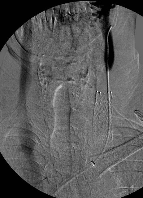
angioplasty was performed at the valvular area (balloon inflated). Then on to evaluate the mid jugular narrowing. Based upon this location, one should consider a few explanations.
1. Perhaps the first angioplasty was extended too high and the normal vein was overstretched and subsequently got scarred down. Not the case this time...
2. Consider that there was compression of the vein by the carotid artery.IVUS showed that this was not the case.
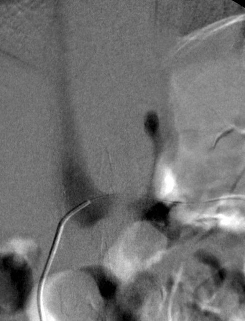
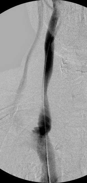
3. Final possibility was muscular compression. The final two images are with the neck rotated, first internally rotated, and the second one externally rotated. You can see that internal rotation results in a phasic complete compression of the vein with no flow at all in the IJV. All the contrast media was directed toward the external jugular vein. . When the head is turned externally, there is no stenosis and there was rapid flow.
Based upon this observation, one can say that this was a phasic narrowing caused by pressure of the muscles of the neck. I would advise no treatment beyond the angioplasty of the confluens' residual tissue narrowing the vein. Dilating muscle compression is unlikely to be helpful. Stenting is a possibility but I prefer NOT to place stents in the jugular except in a few circumstances.