DrSclafani answers some questions

A forum to discuss Chronic Cerebrospinal Venous Insufficiency and its relationship to Multiple Sclerosis.
User avatar
drsclafani
Family Elder
Posts: 3182
Joined: Fri Mar 12, 2010 3:00 pm
Location: Brooklyn, New York
Contact:

Re: DrSclafani answers some questions

Post by drsclafani »

drsclafani wrote:interesting case. I will show the images as soon as they are ready.....

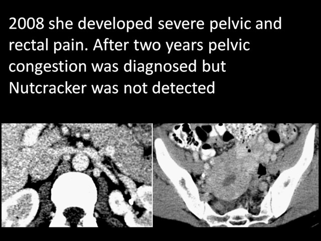
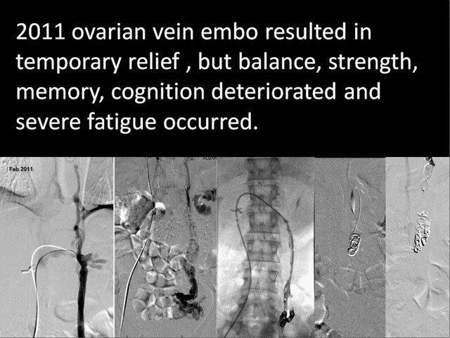
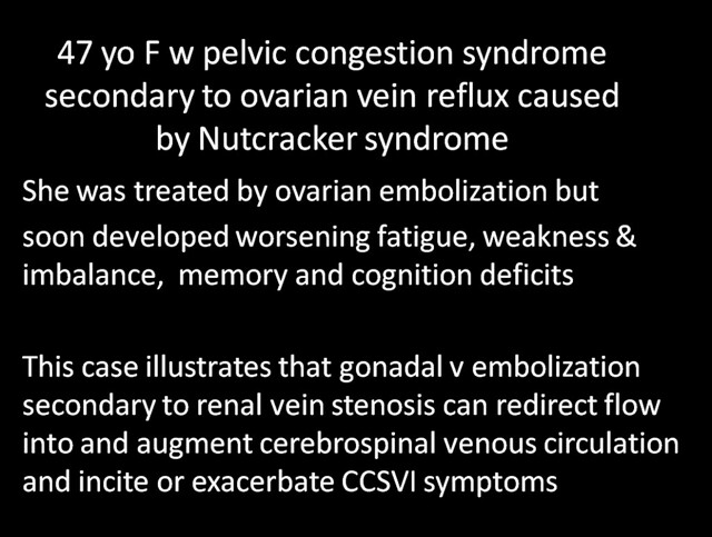
a 47 year old female college professor who was well until four years ago when she began to develop pelvic and rectal pain. After numerous and prolonged testing, she underwent gonadal venography that confirmed the presence of ovarian vein incompetence and varicosities. Pelvic congestion was manifested as well. The ovarian vein was treated by coil and Amplatzer embolization by another interventionalist. Clinical improvements in the pain were short lived. She also began to notice reduced memory, word and name recall and difficulties with multitasking and complex thinking. Balance deteriorated, severe fatigue developed and she became weak in both left upper and lower extremities. Further angiography resulted in coil embolization of the internal iliac vein. Additionally, compression of the left renal vein was identified and a diagnosis of Nutcracker syndrome was made. This was not treated.

She continued to have some relief from the pelvic pain but her new neurological symptoms worsened considerably, resulting in a subjectively diminished quality of life. She sought my consultation, having never seen a neurologist for her problems.

She gave a history of mitral prolapse with mitral regurgitation and unstable knee and poor wound healing. There was no history of multiple sclerosis, optic neuritis or vision disturbance. On physical exam she had a prominent periobital vein, hyperemia of the pharynx, drooping eyelids and double folds in her upper eyelids.She had very lax ligaments in her hands. There was slight weakness in the left hand and the left hip flexor

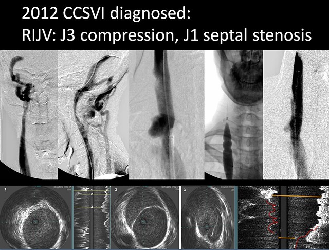
She had a CCSVI protocol neck ultrasound confirmed the presence of two criteria of CCSVI and revealed bilateral IJV reflux in both supine and erect positions in a small left internal jugular vein and a normal right internal jugular vein with a septum creating a infundibulum.

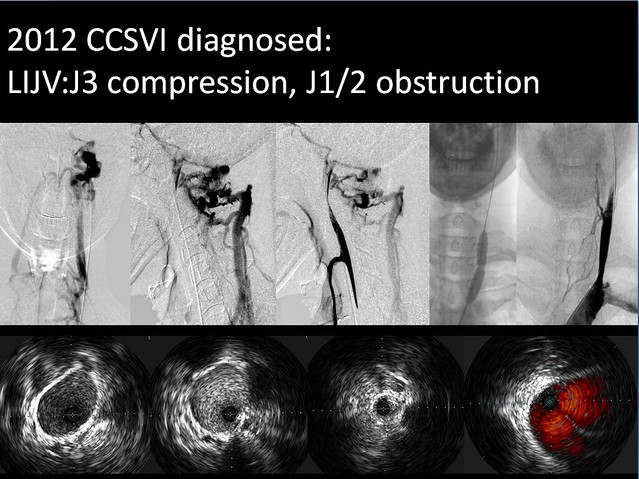
Venography and IVUS confirmed the right jugular vein (IJV) septum and demonstrated a 99% stenosis of the left IJV. Angioplasty of both IJV was performed with satisfactory but not complete dilatation on the left side. The azygous vein was enlarged and there was reflux in the accessory azygous vein.

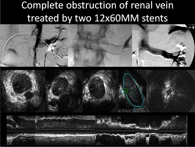
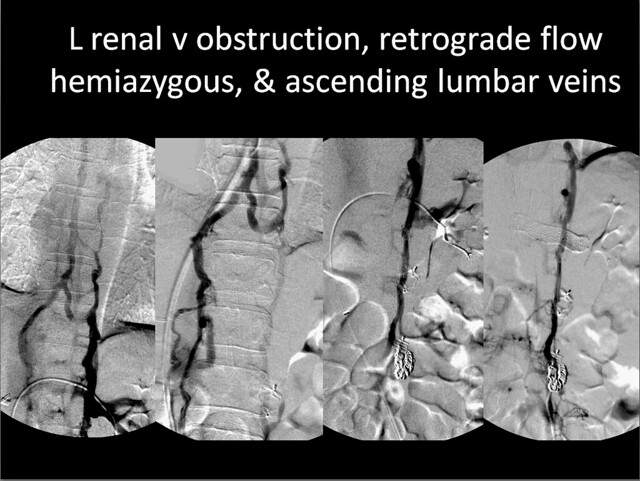
Renal venography showed a classical appearance of the nutcracker. It was very difficult to advance the guidewire or catheter through the obstructed segment that was located at the position of the aorta. Venography revealed total obstruction of the vein. There were no gonadal collaterals but there was complete opacification of the entire vertebral venous system.from T3 to L5. IVUS showed complete compressive obstruction of the vein. After self-expanded stenting between the renal bifurcation and the inferior vena cava, renal blood flow was brisk and the collaterals were no longer seen.
Within 90 minutes she stated that all pelvic pain had resolved and that she felt more alert and clear.

This interesting case shows that in the presence of CCSVI, Nutcracker Syndrome is capable of exacerbating or causing neurological symptoms. With the gonadal veins occluded by coils, renal hemodynamic redirection into the hemiazygous and ascending lumbar veins augmented flow into an already compromised cerebrospinal venous system. The effects were deficits is memory and cognition, balance, strength and the development of a severe fatigue syndrome.

Upon further assessment, Ehlers Danlos syndrome also seems like a real possibility. Do you think these patients are more prone to thrombosis or delayed endothelialization? Should I continue my anticoagulation regimen for an additional few months beyond the standard one month of antithrombin a medications?

I will send on the images as soon as they are ready.
Here are the images.

Image

Image

Image

Image

Image

Image

Image

Image

Any questions?

DrSclafani
Salvatore JA Sclafani MD
Patient contact: ccsviliberation@gmail.com
User avatar
Gardenia
Family Member
Posts: 25
Joined: Wed Jul 06, 2011 2:00 pm

Re: DrSclafani answers some questions

Post by Gardenia »

Doctor
Last year I was treated in the US (I live in Canada) and my azygous vein was found to be the width of a few hairs for a short distance, 80% blockage. I have been in a wheelchair 24/7 since breaking my right leg at 26 since 2002. I was diagnosed with MS at age 23.. Most of my symptoms have pertained to the spine. I did not get stents as the IR was not prepared or rather comfortable with using them. My question is what in your opinion would be the chances that the azygous would narrow up again? The doctor was able to get the vein open wider by about 30% in his estimation. Would it have helped me in anyway or would it be possible to widen the blockage by more than 30%? Your input would be greatly appreciated!
pelopidas
Family Member
Posts: 71
Joined: Mon Sep 05, 2011 6:30 am
Location: Greece

Re: DrSclafani answers some questions

Post by pelopidas »

mtf wrote:Patient: DEIRDRE CARNEGIE

Dx: Multiple Sclerosis, Chronic Cerebrospinal Venous Insufficiency

Ms. Carnegie is a 47 years old patient with Multiple Sclerosis diagnosis since she was 35 years old. However, she presented her symptoms since the age of 17 years old. Mrs. Carnegie symptoms continued to progressed until she presented a progressive and general weakness, in particular sphincter incontinence (urinary and fecal incontinence). In the past she had two Jugular and Azigos vein angioplasty procedures, with excellent results and recovery of lost faculties, in particular the sphincter incontinence.

Months after her last Angioplasty, the symptoms came back, with par aesthesia of the limbs, motor weakness, reduction of the visual acuity, mental confusion and cognitive disorder referred as a foggy thought, but, in particular, the urinary and fecal incontinences. For this reason, the patient came back to Excel Medical Center in Tijuana, where was performed a CT angiography finding a sever occlusion on both jugular veins, particularly the left side.

As a result, she was taken to the Cardio-vascular area, and she has been prepared in order to perform a bilateral jugular angiography, and an angioplasty if it was necessary.

The angiography showed a 99% occlusion of the left jugular vein in the middle third, which could not be corrected by the angioplasty. The right jugular vein presented several severe occlusions in which were corrected by angioplasty using a 12 mm balloon which was inflated at 14 atm's with an excellent results regarding sensory-sensitive symptomatology and partially regarding with motor and cognitive symptomatology. However, the urinary incontinence persisted.

The azygos vein was patent and was not operated.

With these findings, I suggested to the patient to perform a surgical jugular angioplasty by using a vein patch to widen the vein. This treatment named “Jugular Surgical Angioplasty” also known as the “JSA Procedure”, providing a more optimum cerebral venous drainage. The patient accepted. The JSA procedure was performed two days later with excellent results and getting an excellent flow and patency of both vessels.

Remarkably, all the symptomatology that the patient had was almost entirely disappeared in the next 48 hours after the surgical Jugular angioplasty procedure. The most remarkable of these outcomes, is the disappearance of both urinary and fecal incontinences.

The patient was discharged in an excellent status and was advised to take Pradaxar® (Dibigatran) 150 mgs every 12 hrs, ASA Protect 100 mgs ones a day as well as Plavix® (Clopidogrel) tablets ones a day for a 6 weeks period.

She was advised to return to her Neurologist in 4 weeks to be re-evaluated and record the progress or regression of their symptoms.


Dr. S,
What are your comments about this case?

Ernesto L. R. Osuna, M.D., F.A.C.C.

Interventional Cardiologist
[/quote]

It seems very promising. May i ask how big this patch could be and what kind of stenoses could widen? Is it suitable for thrombosed veins?
I think that maybe during this procedure some of the jugular collaterals are ligated and gone for good. This could be a problem if the vein is restenosed again. Or it could be beneficial as in maturation of hypoplastic veins?
Last edited by pelopidas on Mon Feb 13, 2012 10:08 pm, edited 1 time in total.
getafix
User avatar
mo_en
Family Member
Posts: 48
Joined: Thu Dec 09, 2010 3:00 pm
Location: Patras, Greece

Re: DrSclafani answers some questions

Post by mo_en »

Hi Dr S.
I guess the J3 compressions were left untreated. Do you think they could also be the natural outcome of a smaller jugular foramen? Any new ideas about treating this kind of stenoses?
User avatar
HappyPoet
Family Elder
Posts: 1414
Joined: Thu Jul 09, 2009 2:00 pm
Contact:

Re: DrSclafani answers some questions

Post by HappyPoet »

Hi Dr. Sclafani,

RIJV:
1. Considering that J3 wasn't treated, it's unfortunate the waists in the balloon at J1 (septum) couldn't be eradicated as a high degree of stenosis still remains; do you know the highest pressure tried, how many inflations were done, and for how long each inflation lasted?
2. Did the vertebral vein collaterals clear up after angioplasty?
3. In IVUS image #3, is the white line, shown to the right of the middle of the vein, the septum?

LIJV:
1. Considering that J3 wasn't treated, it's unfortunate the waist in the balloon at J1/2 couldn't be eradicated as a degree of stenosis still remains; do you know the highest pressure tried, how many inflations were done, and for how long each inflation lasted?
2. Good to see that many of the vertebral vein collaterals cleared up after the lower jugular angioplasty.
3. What is the significance of the color red in the Iast IVUS image?
Cece
Family Elder
Posts: 9335
Joined: Mon Jan 04, 2010 3:00 pm
Contact:

Re: DrSclafani answers some questions

Post by Cece »

drsclafani wrote:
Image
Did treating the J1 septum improve the appearance of the J3 compression?
Ivus images 2 & 3 are showing the septum? Do you find it easy to distinguish between a septum and a valve?
Image
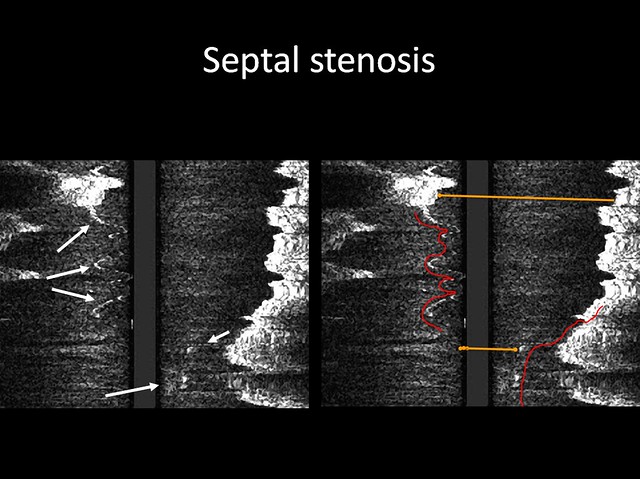
So we are looking at a vertical image of the vein. Anything white is echogenic, meaning there is something there. In the left image, we see the arrows pointing at the white marks. I am not sure how to visualize the septum. In the right image, do the yellow lines indicate the relative width of the vein at the two locations, showing the width is narrower at the lower yellow line. Do those red lines represent one septum? Does the flow go on both sides of the septum? Is one side of the septum in the middle of the red lines, and the other side is closer to us, or to the left of the left red line?
Cece
Family Elder
Posts: 9335
Joined: Mon Jan 04, 2010 3:00 pm
Contact:

Re: DrSclafani answers some questions

Post by Cece »

Image
The first image is the jugular, with very little flow. Are the next two the dural sinus images, shown from the side? There is a lot going on there. The third shows the jugular angioplasty, and the fourth the results, which are not ideal, but improved from the first image.

The first two ivus images show the vein, looking ok, but what is shown in the third ivus image? I find it hard to make out a lumen to the vein. It does not look like the usual valvular blockages that have an obvious vein and a little white line for the valve blockage. Could it be a recanalized thrombosis? My guess on the fourth image is that it's the jugular and the subclavian coming together, each one marked in red?
Cece
Family Elder
Posts: 9335
Joined: Mon Jan 04, 2010 3:00 pm
Contact:

Re: DrSclafani answers some questions

Post by Cece »

Image
That is the best image I've seen of an obstructed renal, and of the improvement once treated.

Why was it necessary to use two stents?
Image
Image by image here.... in the first, we see the catheter going through the renal vein that is completely compressed, so that when the contrast is injected from the right end of the catheter, it flows up the ascending lumbar veins? Which one is the hemiazygous? It looks like there is a stent in place by the fourth image. What is the fourth image showing? Is it showing improvement in flow and reduction in size of those veins once the renal vein has been treated?

I remember being stunned that the renal vein carried as much flow as it did. (500 ml/min, compared to 750 ml/min by the jugulars combined.) All that flow added to the cerebrospinal drainage.
User avatar
ruggierifp
Newbie
Posts: 2
Joined: Sun Aug 22, 2010 2:00 pm

Re: DrSclafani answers some questions

Post by ruggierifp »

; The new procedure is also for CCSVI but it's named the JSA for Jugular Surgical Angioplasty,
Dr. Hernandez has discovered ( This dr's response. Sclafani:


Ernesto, thank you for sharing this very interesting case ... I am thrilled to have this opportunity for patients, whose strictures are not likely to angioplasty. The Patch angioplasty or a bypass graft is a useful tool, for which there is little experience in the internal jugular vein. As we all know, is a unique for its periodic collapse is limited depending on the location of the patient, standing or supine. Will be very helpful to get some followup at short intervals. Would take into account a more long-term anticoagulant therapy. Maintain patency of this vein is very challenging.Please keep us informed.Good luck and we hope to see you on the occasion of the annual meeting of the international society for Neurovascular disease (ISNVD) next week.


For those who are not emotional, this is the video of the intervention


Source http://www.thisisms.com/forum/chronic-c ... -6360.html )

My comments:
In Italy, executed with microsurgery, many interventions on the jugular, n° 40!, by Professor Dr. Dr. SALVATORE Spanish, years or age, 71, of private hospital in Monza (Milan), by replacing with saphenous vein of the leg. To exchange experience on measures to the jugular veins, his phone is 39-335-7033919, his room Polyclinic, 39-039-2810575, his 39-039-2810320. Greetings from Francesco Paolo Ruggieri, present on facebook. E_mail salvatore.spagnolo@policlinico​dimonza.it . Link to facebook, not run by him : https://www.facebook.com/groups/2414988 ... 9700866837 ; ; ; ;


Intervention on the jugular, SONIA CAPOZZOLI, Albanella, SA, on Facebook, pre-operative, 6 months have forsaken therapies for MS and detoxicate the organism, with CLADOPHYLL and 250 g of OLIVE OIL, BIODYNAMIC, GOLDEN YELLOW and thick, TAKEN DURING the DAY, for ABOUT 5 MONTHS, result JUN VEINS. INNER DIAMETER of 14 mm THICK, and analysis, perfect, with VERY QUICK HEALING and STAND by 12:0 am FROM intervention and, AFTER ABOUT 25-30 days, DO go, RACE and WALK, a 5 km ROUTE!
User avatar
drsclafani
Family Elder
Posts: 3182
Joined: Fri Mar 12, 2010 3:00 pm
Location: Brooklyn, New York
Contact:

Re: DrSclafani answers some questions

Post by drsclafani »

Gardenia wrote:Doctor
Last year I was treated in the US (I live in Canada) and my azygous vein was found to be the width of a few hairs for a short distance, 80% blockage. I have been in a wheelchair 24/7 since breaking my right leg at 26 since 2002. I was diagnosed with MS at age 23.. Most of my symptoms have pertained to the spine. I did not get stents as the IR was not prepared or rather comfortable with using them. My question is what in your opinion would be the chances that the azygous would narrow up again? The doctor was able to get the vein open wider by about 30% in his estimation. Would it have helped me in anyway or would it be possible to widen the blockage by more than 30%? Your input would be greatly appreciated!
The answer to this question really depends upon the location of the stenosis.Some of these narrowings depend upon the phase of respiration rather than being fixed. You can dilate but the narrowing comes back no matter what you do if the pictures are taken in expiration rather than inspiration. Going from 80% to 50% is not sufficient if this is a real stenosis. I havent seen problems with stents in the azygous vein in the few patients who have come to see me after being treated by someone else.
Salvatore JA Sclafani MD
Patient contact: ccsviliberation@gmail.com
User avatar
drsclafani
Family Elder
Posts: 3182
Joined: Fri Mar 12, 2010 3:00 pm
Location: Brooklyn, New York
Contact:

Re: DrSclafani answers some questions

Post by drsclafani »

pelopidas wrote:
mtf wrote:Patient: DEIRDRE CARNEGIE

Dx: Multiple Sclerosis, Chronic Cerebrospinal Venous Insufficiency

Ms. Carnegie is a 47 years old patient with Multiple Sclerosis diagnosis since she was 35 years old. However, she presented her symptoms since the age of 17 years old. Mrs. Carnegie symptoms continued to progressed until she presented a progressive and general weakness, in particular sphincter incontinence (urinary and fecal incontinence). In the past she had two Jugular and Azigos vein angioplasty procedures, with excellent results and recovery of lost faculties, in particular the sphincter incontinence.

Months after her last Angioplasty, the symptoms came back, with par aesthesia of the limbs, motor weakness, reduction of the visual acuity, mental confusion and cognitive disorder referred as a foggy thought, but, in particular, the urinary and fecal incontinences. For this reason, the patient came back to Excel Medical Center in Tijuana, where was performed a CT angiography finding a sever occlusion on both jugular veins, particularly the left side.

As a result, she was taken to the Cardio-vascular area, and she has been prepared in order to perform a bilateral jugular angiography, and an angioplasty if it was necessary.

The angiography showed a 99% occlusion of the left jugular vein in the middle third, which could not be corrected by the angioplasty. The right jugular vein presented several severe occlusions in which were corrected by angioplasty using a 12 mm balloon which was inflated at 14 atm's with an excellent results regarding sensory-sensitive symptomatology and partially regarding with motor and cognitive symptomatology. However, the urinary incontinence persisted.

The azygos vein was patent and was not operated.

With these findings, I suggested to the patient to perform a surgical jugular angioplasty by using a vein patch to widen the vein. This treatment named “Jugular Surgical Angioplasty” also known as the “JSA Procedure”, providing a more optimum cerebral venous drainage. The patient accepted. The JSA procedure was performed two days later with excellent results and getting an excellent flow and patency of both vessels.

Remarkably, all the symptomatology that the patient had was almost entirely disappeared in the next 48 hours after the surgical Jugular angioplasty procedure. The most remarkable of these outcomes, is the disappearance of both urinary and fecal incontinences.

The patient was discharged in an excellent status and was advised to take Pradaxar® (Dibigatran) 150 mgs every 12 hrs, ASA Protect 100 mgs ones a day as well as Plavix® (Clopidogrel) tablets ones a day for a 6 weeks period.

She was advised to return to her Neurologist in 4 weeks to be re-evaluated and record the progress or regression of their symptoms.


Dr. S,
What are your comments about this case?

Ernesto L. R. Osuna, M.D., F.A.C.C.

Interventional Cardiologist
It seems very promising. May i ask how big this patch could be and what kind of stenoses could widen? Is it suitable for thrombosed veins?
I think that maybe during this procedure some of the jugular collaterals are ligated and gone for good. This could be a problem if the vein is restenosed again. Or it could be beneficial as in maturation of hypoplastic veins?[/quote]

the video looked like they extracted thrombus. I agree with your concerns about collaterals. I also worry about reocclusion because of the intermittent nature of jugular flow.

we shall see, eh pelopidas!
Salvatore JA Sclafani MD
Patient contact: ccsviliberation@gmail.com
User avatar
drsclafani
Family Elder
Posts: 3182
Joined: Fri Mar 12, 2010 3:00 pm
Location: Brooklyn, New York
Contact:

Re: DrSclafani answers some questions

Post by drsclafani »

mo_en wrote:Hi Dr S.
I guess the J3 compressions were left untreated. Do you think they could also be the natural outcome of a smaller jugular foramen? Any new ideas about treating this kind of stenoses?

These stenoses are the result of compression between muscles and the vertebra at that level. Often this narrowing opens up in extension of the neck. It did not open up in this case. It is my practice to treat other stenoses first and see how the patient responds. if no good response, then patient and i confer about stenting that area. I really do not want to stent unless really proven to be necessary and worth effort and risk. So we shall see h ow the patient does.

I think you can see that the jugular foramen is large because there is no narrowing of the contrast column as the vein travels through the bony canal. There is nothing minimally invasive that can be done for a small canal. the patient will have to rely upon condylar emissary vein for collateral flow. In such cases I would want to see that there are no obstructions of the outflow vein for the condylar vein, something i havent thought about until you asked this question.

what a team! Thanks
Salvatore JA Sclafani MD
Patient contact: ccsviliberation@gmail.com
Cece
Family Elder
Posts: 9335
Joined: Mon Jan 04, 2010 3:00 pm
Contact:

Re: DrSclafani answers some questions

Post by Cece »

With ISNVD coming up, I am glad we have you there representing us, and representing the need for as complete a CCSVI treatment as possible! Using IVUS is about being thorough and being safe. Treating renal stenosis is about doing everything that can be done to improve cerebrospinal flow. Checking the veins into which the condylar emissary vein flows? Thorough, thorough, thorough. And it makes a difference for the patients.
User avatar
drsclafani
Family Elder
Posts: 3182
Joined: Fri Mar 12, 2010 3:00 pm
Location: Brooklyn, New York
Contact:

Re: DrSclafani answers some questions

Post by drsclafani »

HappyPoet wrote:Hi Dr. Sclafani,

RIJV:
1. Considering that J3 wasn't treated, it's unfortunate the waists in the balloon at J1 (septum) couldn't be eradicated as a high degree of stenosis still remains; do you know the highest pressure tried, how many inflations were done, and for how long each inflation lasted?
2. Did the vertebral vein collaterals clear up after angioplasty?
3. In IVUS image #3, is the white line, shown to the right of the middle of the vein, the septum?

LIJV:
1. Considering that J3 wasn't treated, it's unfortunate the waist in the balloon at J1/2 couldn't be eradicated as a degree of stenosis still remains; do you know the highest pressure tried, how many inflations were done, and for how long each inflation lasted?
2. Good to see that many of the vertebral vein collaterals cleared up after the lower jugular angioplasty.
3. What is the significance of the color red in the Iast IVUS image?

The J3 stenoses are compressions against the spine. they may be phasic or fixed. These appeared fixed. Treatment would probably require stenting so I decided to treat the other stenoses and see what happens.
Those are not vertebral collaterals but condylar vein collaterals
The red line is outlining the septum.

The right sided septum seems to have responded to moderately high pressure 13 Atm and lost its waist. I was satisfied that collaterals were not significant after angioplasty. we will see if this remains so. I am concerned that septum seems to recur often.
Salvatore JA Sclafani MD
Patient contact: ccsviliberation@gmail.com
User avatar
drsclafani
Family Elder
Posts: 3182
Joined: Fri Mar 12, 2010 3:00 pm
Location: Brooklyn, New York
Contact:

Re: DrSclafani answers some questions

Post by drsclafani »

Cece wrote:
drsclafani wrote:
Image
Did treating the J1 septum improve the appearance of the J3 compression?
Ivus images 2 & 3 are showing the septum? Do you find it easy to distinguish between a septum and a valve?
No, the J3 segment was still stenotic

No, sometimes it is difficult to differentiate them. i am pretty sure this is a septum
Image
So we are looking at a vertical image of the vein. Anything white is echogenic, meaning there is something there. In the left image, we see the arrows pointing at the white marks. I am not sure how to visualize the septum.
the arrow is pointing to the septum
In the right image, do the yellow lines indicate the relative width of the vein at the two locations, showing the width is narrower at the lower yellow line. Do those red lines represent one septum? Does the flow go on both sides of the septum? Is one side of the septum in the middle of the red lines, and the other side is closer to us, or to the left of the left red line?
It is really difficult to completely understand what we are looking at. To me, i see the septum attached to one wall at the top, and as we go down the septum moves toward the opposite wall. lower still there is some echogenic material on the other side. Is it spiraling down around the catheter?? Is this the septum attached to the opposoite walls? Difficult to tell perfectly because of inexperience and the lack of pathological followups.
Salvatore JA Sclafani MD
Patient contact: ccsviliberation@gmail.com
Post Reply
  • Similar Topics
    Replies
    Views
    Last post

Return to “Chronic Cerebrospinal Venous Insufficiency (CCSVI)”