DrSclafani answers some questions

A forum to discuss Chronic Cerebrospinal Venous Insufficiency and its relationship to Multiple Sclerosis.
Cece
Family Elder
Posts: 9335
Joined: Mon Jan 04, 2010 3:00 pm
Contact:

Re: DrSclafani answers some questions

Post by Cece »

drsclafani wrote:The obstruction is actually visible on these images. You have to discriminate the contrast in the proximal vein from the contrast media that is in the vein below the obstruction

Image

When looking again, and with it labelled and an arrow, I can see the stenosis as a darker narrowing funnel above and within the lighter contrast.
David1949
Family Elder
Posts: 927
Joined: Mon Aug 23, 2010 2:00 pm
Contact:

Re: DrSclafani answers some questions

Post by David1949 »

Cece wrote:pssst anyone else want to join in?

Image
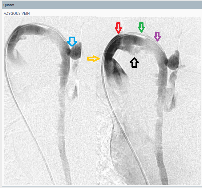
I added arrows to be more clear.
Blue arrow is pointing at the "odd attachment." What is that?
Yellow arrow is pointing at what looks like a narrowing before we're even into the azygous proper.
Red arrow is pointing at what seems to be a shift from darker contrast to medium contrast. Green arrow is where it shifts again from medium contrast to light contrast. Purple arrow is the light contrast. I would have guessed compression at the purple arrow if it weren't for the mention of aneurysms.
Black arrow looks like a possible aneurysm. But more than one aneurysm was mentioned...
I haven't learned "Doctor speak" yet. But undaunted by my lack of comprehension let me chime in anyway. It was mentioned that the odd protrusion is due to a perforation. Has this patient had venography before? If so, is it possible that the tip of the wire could have caused a perforation? The wire has a very small diameter and looks almost sharp on the end.

OOPS! Looks like my questions were already answered. Sorry about that!
Last edited by David1949 on Sat Apr 14, 2012 5:04 pm, edited 2 times in total.
David1949
Family Elder
Posts: 927
Joined: Mon Aug 23, 2010 2:00 pm
Contact:

Re: DrSclafani answers some questions

Post by David1949 »

Speaking of wires, is it possible that the tip of the wire could scratch the wall of the vein and cause blod clots or intimal hyperplasia?

When arteries are treated by angioplasty do they have the same problems with blod clots and intimal hyperplasia?
User avatar
drsclafani
Family Elder
Posts: 3182
Joined: Fri Mar 12, 2010 3:00 pm
Location: Brooklyn, New York
Contact:

Re: DrSclafani answers some questions

Post by drsclafani »

David1949 wrote:Speaking of wires, is it possible that the tip of the wire could scratch the wall of the vein and cause blod clots or intimal hyperplasia?

When arteries are treated by angioplasty do they have the same problems with blod clots and intimal hyperplasia?
david, guidewires are generally curved or have floppy tips so that as they touch something they buckle over and do not lead with their pointy tip. But it is definitely possible to perforate a blood vessel, vein more easily than an artery. It is not so likely that they will cause clots or intimal hyperplasia so easily.

it is possible to feel the wire touching the wall of the blood vessel as you move it. it can be a subtle but an important skill
Salvatore JA Sclafani MD
Patient contact: ccsviliberation@gmail.com
User avatar
drsclafani
Family Elder
Posts: 3182
Joined: Fri Mar 12, 2010 3:00 pm
Location: Brooklyn, New York
Contact:

Re: DrSclafani answers some questions

Post by drsclafani »

Here is another interesting case of damage to an azygous vein. A 48 year old female with major complaints of cog-fog, vertigo, imbalance, numbness in feet, hands and back, pain the the legs, weakness in the legs right atm and back, sometimes with spasticity and periodic urinary symptoms Imbalance required the use of a cane for walking.
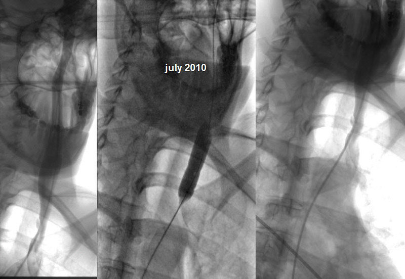
this patient was treated in Europe. Image
The azygous vein was not patent and it took considerable effort to open this apparently occluded or hypopplastic vein.


Image
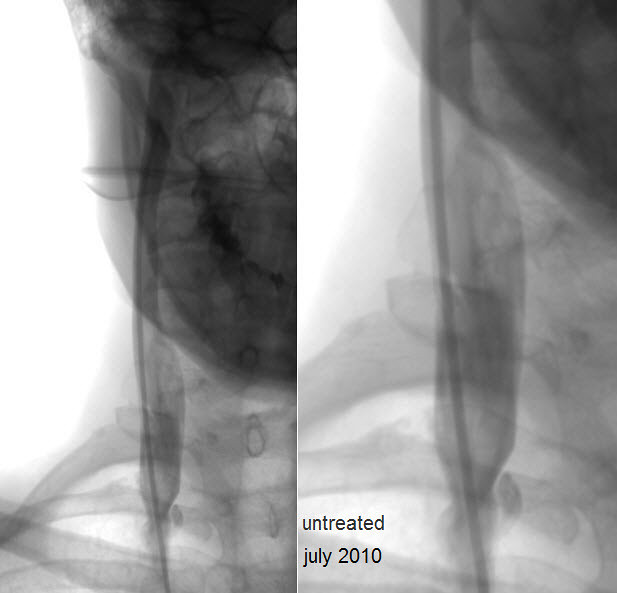
The left internal jugular vein was thought to be 30% ste4nosed and it was treated by a balloon that seems undersized balloon


Image
The right internal jugular vein was deemed to have only a 20% stenosis and was not treated


This patient had improvements in balance, fatigue, cogfog and numbness. However she had relapse about six xmonths after treatment and over the subsequent eight months she gradually lost all improvements. She came for consultation. She had not had iliac, renal or lumbar angiography and I believed that the jugular veins were not satisfactorily treated.

I will show you how i approached this procedure after you have had some time to look this over
Salvatore JA Sclafani MD
Patient contact: ccsviliberation@gmail.com
User avatar
Robnl
Family Elder
Posts: 668
Joined: Sun Nov 08, 2009 3:00 pm
Contact:

Re: DrSclafani answers some questions

Post by Robnl »

Hi Doc,
The left internal jugular vein was thought to be 30% stenosed and it was treated by a balloon that seems undersized balloon
- Is 30% enough to balloon? A.k.a. from what percentage do you think it's useful to balloon?
- How do you 'see' that an undersized balloon is used?
User avatar
mo_en
Family Member
Posts: 48
Joined: Thu Dec 09, 2010 3:00 pm
Location: Patras, Greece

Re: DrSclafani answers some questions

Post by mo_en »

Hi Dr S, i wish you a quick recovery and a stormy comeback.

In the case above, in the azygos image far right, the catheter is outside the opacified portion of the vein. Is this an artifact or is the vein divided by something along its course?

In the jugular veins the protruding valves seem rather obvious, especially in the right JV. I bet the percentages of occlusion were significantly higher.
Last edited by mo_en on Tue Apr 17, 2012 5:57 am, edited 1 time in total.
User avatar
drsclafani
Family Elder
Posts: 3182
Joined: Fri Mar 12, 2010 3:00 pm
Location: Brooklyn, New York
Contact:

Re: DrSclafani answers some questions

Post by drsclafani »

Robnl wrote:Hi Doc,
The left internal jugular vein was thought to be 30% stenosed and it was treated by a balloon that seems undersized balloon
- Is 30% enough to balloon? A.k.a. from what percentage do you think it's useful to balloon?
- How do you 'see' that an undersized balloon is used?
This is a good question. The conventional thought revolves around 50% stenosis. But this was an arbitrary determination in arteries. But such percentages seem to be associated IN ARTERIES with pressure gradients where the pressure drops on the distal side of the stenosis. Moreover in arteries you can chemically exaggerate the drop in pressures by administering certain medications that can resemble the effects of stress so that we have the ability to correlate pressure with degree of stenosis.

Veins are much different because they are about flow not pressure. gradients across stenoses can be as little as 3mm and be significant. Also with significant flow veins expand greatly unlike arteries that are relatively stable in their size.
When flow increases and size increases the stenoseis is exaggerated and the stenosis appears to be a greater percentage.

So it is not so simple. I use secondary signs of venous obstruction, including large collateral veins, slow flow, reflux or to/from movement of the contrast media and stasis of the contrast media.

and i use IVUS which is overwhelmingly better at determining constriction of the dimensions of a tube. while we can see a slight narrowing in one direction on venogram, the irregular shapes of veins may mislead us in that one view. Diameter measurements do not reflux the dimensions of a vein like circumference measurements do. IVUS gives us circumference measurements that are accurate to a tenth of a square millimeter. Also circumference is calculated as a square of the radius which magnifies any differences compared to measurements using diameter.

So lets look at the images used by the prior physician and compare to the images i create to make this determination.
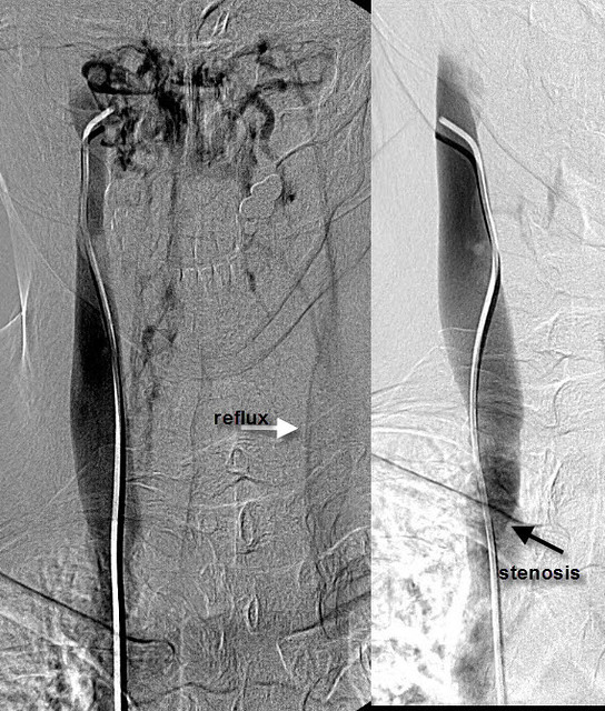
Image
you saw these two images on the far left when i presented the case. I thought it was more significant that the prior physician and I would have treated it if it were my patient at that time. The next image to the right shows my venogram done this month. You can see that there is not much difference from the first two in terms of the stenosis.

The next two images are IVUS images of the area of the stenosis. The upper one shows the valvular tissue with small white arrows indicating the opening of the valve. THIS is the stenosis as all the blood above must travel through this constriction of the valve. The lower images is the SAME image with blue outlining the dimensions of the vein and the green outlining the dimensions of the opening of the valve. It may be difficult to read, but below this are m easurements of the circumferences of the two and one can see that the percentage difference of the two circumferences is close to 70% and this would lead to no disagreement by anyone. Moreover this is NOT subjective, it is inarguable measured fact. The last two images show the constrictions in the balloons during dilation and the end result with the vein wide open, very rapid flow and a successful treatment.

That is how i do it.

i have to go to work, but i will explain why i think the left balloon was undersized. and how i determine what size balloon to use.

Thanks for asking
DrS
Salvatore JA Sclafani MD
Patient contact: ccsviliberation@gmail.com
Cece
Family Elder
Posts: 9335
Joined: Mon Jan 04, 2010 3:00 pm
Contact:

Re: DrSclafani answers some questions

Post by Cece »

drsclafani wrote:It may be difficult to read, but below this are m easurements of the circumferences of the two and one can see that the percentage difference of the two circumferences is close to 70% and this would lead to no disagreement by anyone. Moreover this is NOT subjective, it is inarguable measured fact.
That is quite a difference between the estimate by the original treating IR of the stenosis being 20% and the precise measurement on IVUS showing it to be 70%.

No questions from me on that.
i have to go to work, but i will explain why i think the left balloon was undersized. and how i determine what size balloon to use.
I don't see waisting on the balloon in the left jugular. When looking at the final outcome image, which looks much improved from the first image, is there a bulge remaining at the area of the valves? What was the measurement of this vein by IVUS, and what was the size of the balloon used in the original procedure?

I am worried about the damage to the azygous vein, as mentioned:
Here is another interesting case of damage to an azygous vein
Did it occlude?
I suppose even if it occluded, the ascending portion was already occluded or hyperplastic in the initial pre-treatment image. It is different than losing a functioning azygous vein. But, still upsetting.
David1949
Family Elder
Posts: 927
Joined: Mon Aug 23, 2010 2:00 pm
Contact:

Re: DrSclafani answers some questions

Post by David1949 »

May I ask if the patient saw improvements in her symptoms after you treated her?
User avatar
drsclafani
Family Elder
Posts: 3182
Joined: Fri Mar 12, 2010 3:00 pm
Location: Brooklyn, New York
Contact:

Re: DrSclafani answers some questions

Post by drsclafani »

mo_en wrote:Hi Dr S, i wish you a quick recovery and a stormy comeback.

In the case above, in the azygos image far right, the catheter is outside the opacified portion of the vein. Is this an artifact or is the vein divided by something along its course?
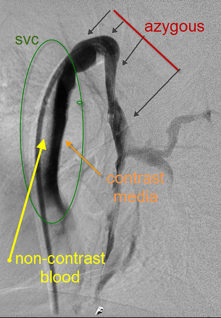
this annotated image should explain things

Image

The reason that the catheter looks like it is outside the vessel is that the vessel is not outlined by contrast media. First, lets review the anatomy. There are two vessels on this image: the upper arrows point to the azygous vein and the area of green circle points to the other vessel, the superior vena cava into which the azygous vein drains. The superior vena cava is not completely filled with contrast media. Why? The patient is lying on her back. Because the dye is heavier than blood, it layers below the blood, sort of like oil and water. So you see the upper part (yellow) filled with blood and the lower part (orange) filled with contrast. The catheter is just in the part of the superior vena cava that is not opacified by contrast media.
Salvatore JA Sclafani MD
Patient contact: ccsviliberation@gmail.com
User avatar
drsclafani
Family Elder
Posts: 3182
Joined: Fri Mar 12, 2010 3:00 pm
Location: Brooklyn, New York
Contact:

Re: DrSclafani answers some questions

Post by drsclafani »

mo_en wrote:
In the jugular veins the protruding valves seem rather obvious, especially in the right JV. I bet the percentages of occlusion were significantly higher.
I agree with you about the abnormality but we do not consider it percentage of occlusion since the veins are not occluded. We talk about percentage of stenosis.
Salvatore JA Sclafani MD
Patient contact: ccsviliberation@gmail.com
User avatar
1eye
Family Elder
Posts: 3780
Joined: Wed Mar 17, 2010 3:00 pm
Location: Kanata, Ontario, Canada
Contact:

Re: DrSclafani answers some questions

Post by 1eye »

Yeah I thought there was a membrane there, but the ink shows it very clearly causes a problem. Hardly visible on the IVUS.
This unit of entertainment not brought to you by FREMULON.
Not a doctor.
"I'm still here, how 'bout that? I may have lost my lunchbox, but I'm still here." John Cowan Hartford (December 30, 1937 – June 4, 2001)
User avatar
drsclafani
Family Elder
Posts: 3182
Joined: Fri Mar 12, 2010 3:00 pm
Location: Brooklyn, New York
Contact:

Re: DrSclafani answers some questions

Post by drsclafani »

Robnl wrote:Hi Doc,- How do you 'see' that an undersized balloon is used?
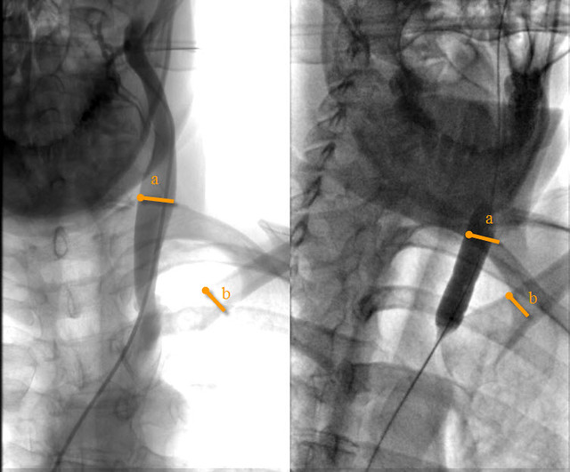
look at this image
Image

this is a composite of the jugular vein stenosis and the inflated balloon used.

Two orange lines are measurements. A= the vein and the balloon and B= the same area of the clavicle. The clavicle was measured to determine which image has more magnification. you can see that the right sided image of the clavicle is larger than the one on the left. This indicates that there is more magnification on the right image. So when we measure the balloon size and we see that it is smaller than the image of the vein on the left, we know that the balloon is undersized. This despite the fact that the image on the right shows that there is more magnification in the right image.
Last edited by drsclafani on Tue Apr 17, 2012 8:48 pm, edited 1 time in total.
Salvatore JA Sclafani MD
Patient contact: ccsviliberation@gmail.com
User avatar
drsclafani
Family Elder
Posts: 3182
Joined: Fri Mar 12, 2010 3:00 pm
Location: Brooklyn, New York
Contact:

Re: DrSclafani answers some questions

Post by drsclafani »

Cece wrote:When looking at the final outcome image, which looks much improved from the first image, is there a bulge remaining at the area of the valves? What was the measurement of this vein by IVUS, and what was the size of the balloon used in the original procedure?
There is a slight bulge still persisting. Not much I could do about that but i do not think it is significant. it was reported that a 12 mm balloon was used in 2010. I used a 16 mm balloon at 20 atmospheres. I used an 18 mm balloon on the right jugular vein.
cece wrote:I am worried about the damage to the azygous vein, as mentioned:
Did it occlude?
I suppose even if it occluded, the ascending portion was already occluded or hyperplastic in the initial pre-treatment image. It is different than losing a functioning azygous vein. But, still upsetting.
correction: it is hypoplastic.

This vein was heroically worked on. However. while it was opened, one can see extravasation of contrast media to the right of the vein. I suspect that it wasnt worth the effort as i have not been very impressed with trying to open hypoplastic veins. Yes it was occluded on the venogram i performed. I did not get heroic this time around.
Salvatore JA Sclafani MD
Patient contact: ccsviliberation@gmail.com
Post Reply
  • Similar Topics
    Replies
    Views
    Last post

Return to “Chronic Cerebrospinal Venous Insufficiency (CCSVI)”