Yes, well. There was a reason that i mentioned the PPMS. As we know, ppms is supposed to have a higher incidence of azygous disease.
The internal jugular veins were imaged. The right side was normal by venography and IVUS.
Here is an image of the left internal jugular vein.
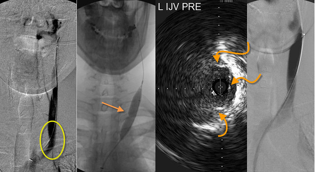
There is a stenosis at the confluens on the venogram (yellow oval). Note near the skull the collateral veins in the perivertebral plexus. There is thickened valvular material (curved orange arrows) on the IVUS that points to the cause of the narrowing of the LIJV as being valvular in nature. After angioplasty (orange arrow) the lumen looked great, and flow was improved.
Also it has been said to be uncommon to have only one vein obstruction in ccsvi.
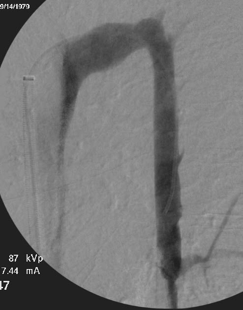
So with one normal ijv and one abnormal ijv, we have to look carefully at the aygous vein. this Azygous is the mother of all azygous. on IVUS it measured 18 mm. well beyond the usual range of 8-12 mm
I can think of several reasons for enlargement of the azygous vein
1. obstruction of both jugular veins
2. obstruction of the left renal vein
3. obstruction of the left iliac vein
4 high flow arteriovenous fistula that drains into the aygous vein
5. obstruction of the outflow drainage of the aygous vein.
some of the group answere my question, talking about the large expansion of the arch and dr sossi talks about the ascending azygous also being enlarge. The ascending vessel measured about 9.5 mm. This is somewhat large but nowhere near the dimension of the arch.
But do we see a beak like narrowing at the orifice of the vein, to the left in the image? No. Do we see filling defects in the contrast column? no.
So lets proceed to look at the pelvis. Obstructions of flow from the leg into the inferior vena cava, the large main vein that carries blood from the lower half of the body into the heart, force that blood to seek alternate pathways back to the heart. These pathways cross from left to right through collaterals in the pelvis and through the ascending lumbar vein into the hemiazygous vein and finally into the aygous vein.
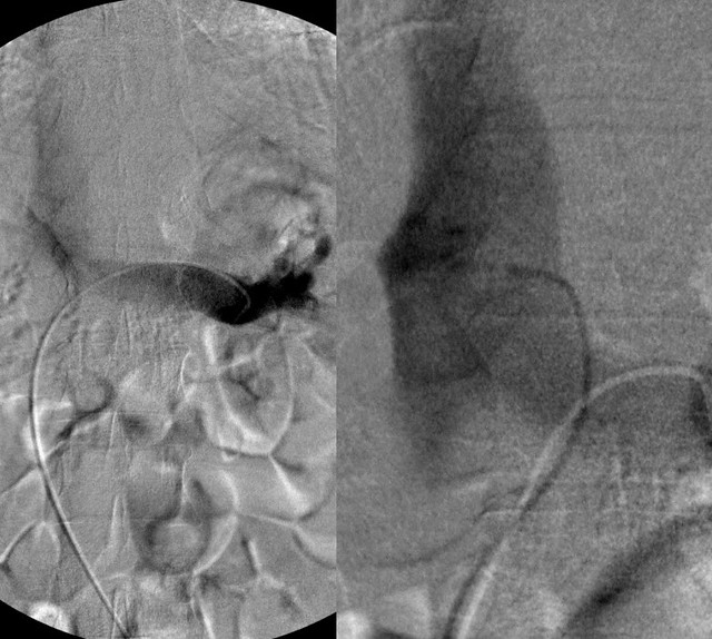
in this patient we see a stenosis of the external iliac vein

This was treated by balloon angioplasty. the results are good. Note also on the film on the right that there is a duplication of the common iliac vein. I did not dilate that segment.
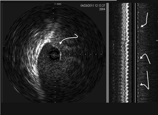
The cause of the stenosis is nicely shown by IVUs to be a septum in the vein (small curved arrows)
Another cause of enlargement of the aygous vein is drainage of the left kidney's venous blood into the azygous vein. This can happen when there is an obstruction of the renal vein by stenosis or occlusion. Then blood can shunt up the ascending lumbar vein, into the hemiazygous vein and into the azygous vein. The left kidney accounts for at least 10% of blood flow.
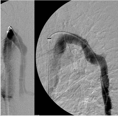
The renal venogram shows nonocclusion of the left renal vein.
i will review the azygous vein tomorrow