DrSclafani answers some questions

A forum to discuss Chronic Cerebrospinal Venous Insufficiency and its relationship to Multiple Sclerosis.
User avatar
drsclafani
Family Elder
Posts: 3182
Joined: Fri Mar 12, 2010 3:00 pm
Location: Brooklyn, New York
Contact:

Post by drsclafani »

Cece wrote:
drsclafani wrote:Image
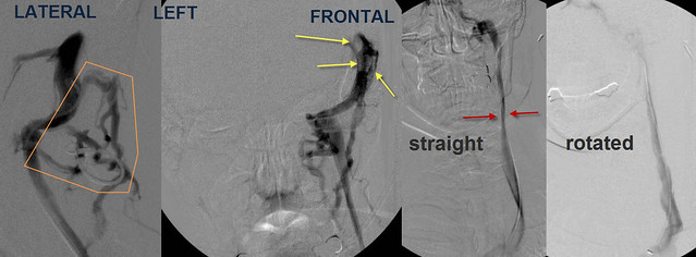
Selective catheterization and venography of the left sigmoid sinus showed that unlike the right side extensive collateral veins were noted in the back of the neck (orange) Also noted were filling defects, consistent with thrombus, in the sinuses. Frontal venogram of the L IJV showed narrowing in J2 (red arrows) similar to the right side. Again, upon rotation of the neck, flow was improved and diameter was normal.
Will there be any treatment for the patient's sinus thrombosis?
i think that this was old clot. i didnt think that it would respond to thrombolytic agents. a mechanical thrombectomy device might have been helpful but i did not think it would go through the jugular bulb and canal. So the best i could do was flush the clots, hopefully down the other sigmoid sinus which was already opened and the jugular had great flow on that side. some of the clot came down the left and some of that got hung up in J3. A large bore open mouthed catheter was used to suck out the clot. the tissue was very hard and firm suggesting that it was an old clot. Perhaps it was a residual of the prior ccsvi procedure
Did the patient have any symptoms of sinus thrombosis or a guess as to when it occurred in the patient history?
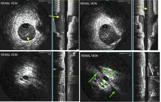
this was an incomplete thrombus. such things can cause symptoms simular to ccsvi. (well known)
Was there any external/physical appearance to the neck to suggest the problematic muscles? Was CTOS discussed? Could neck stretches help these muscles loosen up?
no normal looking neck. at this time i am not convinced about ctos. i want to treat first and see if there is improvement.

So we will treat the renal vein first.
Cece
Family Elder
Posts: 9335
Joined: Mon Jan 04, 2010 3:00 pm
Contact:

Post by Cece »

drsclafani wrote:Thanks for asking
yes i have now two cases. both have azygous disease.
i think that these are truncal malformations in many circumstances wheree the veins of the fetus malform during transition between the fetal veins and the adult veins.
Is there anyone reading this who had his renal vein treated by Dr. McGuckin or Dr. Kirsch but did NOT have his azygous treated?

It may be that in patients with azygous disease, also caused by truncal malformation, the incidence of truncal malformation renal stenosis is higher than in the regular population.

Dr. Siskin used the fact that iliac disease was not found in MS patients to any higher degree than the the incidence in the regular population as justification for not interrogating the iliac. (see ccsvi.org video for his discussion of this)

I imagine the same argument might be applied for whether or not to look at the renal vein, especially if something is only found about once every 75 - 100 patients. I'd disagree with such an argument, of course.
User avatar
drsclafani
Family Elder
Posts: 3182
Joined: Fri Mar 12, 2010 3:00 pm
Location: Brooklyn, New York
Contact:

Post by drsclafani »

Cece wrote:
drsclafani wrote:Thanks for asking
yes i have now two cases. both have azygous disease.
i think that these are truncal malformations in many circumstances wheree the veins of the fetus malform during transition between the fetal veins and the adult veins.
Is there anyone reading this who had his renal vein treated by Dr. McGuckin or Dr. Kirsch but did NOT have his azygous treated?

It may be that in patients with azygous disease, also caused by truncal malformation, the incidence of truncal malformation renal stenosis is higher than in the regular population.

Dr. Siskin used the fact that iliac disease was not found in MS patients to any higher degree than the the incidence in the regular population as justification for not interrogating the iliac. (see ccsvi.org video for his discussion of this)

I imagine the same argument might be applied for whether or not to look at the renal vein, especially if something is only found about once every 75 - 100 patients. I'd disagree with such an argument, of course.
i think that dr siskin missed the point. We do not look for may thurner syndrome in ccsvi because it is more frequent in patients with ccsvi; we look for it because its presence is hemodynamically more significant in patients with ccsvi. This was the point that Robbie galeotti brought home to me.

it will be really telling if my nonresponding to angioplasty patient responds to correcting renal blood flow
Cece
Family Elder
Posts: 9335
Joined: Mon Jan 04, 2010 3:00 pm
Contact:

Post by Cece »

drsclafani wrote:i think that dr siskin missed the point. We do not look for may thurner syndrome in ccsvi because it is more frequent in patients with ccsvi; we look for it because its presence is hemodynamically more significant in patients with ccsvi. This was the point that Robbie galeotti brought home to me.
This is succinctly said.

I knew Nunzio had posted about the renal vein before, here is a post from him with an image from Dr. Galeotti's presentation illustrating some of this.
www.thisisms.com/ftopicp-159910.html#159910
Dr. Galeotti's percentage was 5% of MS patients who had renal stenosis.
it will be really telling if my nonresponding to angioplasty patient responds to correcting renal blood flow
Now there's a cliffhanger! :)
I hope treatment is soon and successful.
Cece
Family Elder
Posts: 9335
Joined: Mon Jan 04, 2010 3:00 pm
Contact:

Post by Cece »

drsclafani wrote:One notes in (C) a stenosis in the J2 segment with collaterals bridging the area of stenosis and entering the inominate vein. Generally, this would be considered a hypoplastic segment. However I have started to see that muscular compression can cause this appearance.
Typically, with muscular compression, is it symmetrical like this so that there is muscular compression on both the RIJV and the LIJV? That was a surprise to me, that this patient had it on both sides.
Cece
Family Elder
Posts: 9335
Joined: Mon Jan 04, 2010 3:00 pm
Contact:

Post by Cece »

IVUS showed that there was echogenic material in the sigmoid sinus and jugular bulb (orange stars Image A&B). In the upper jugular vein in J3 IVUS showed a thrombus (orange arrows). More distally at the confluens a septum was seen narrowing the jugular lumen (curved orange arrows)
If you had not had IVUS and/or you had not entered the sigmoid sinus, would you still have diagnosed the thrombus and treated it the same way?
Cece
Family Elder
Posts: 9335
Joined: Mon Jan 04, 2010 3:00 pm
Contact:

Post by Cece »

I am posting questions separately, I figure it is easier to answer that way....
In August 2010 she was treated by angioplasty by an established American Group practice by angioplasty of the jugular veins. She immediately improved. Notably her vision was "perfect". Sadly, the improvement only lasted two weeks.
What anticoagulation regimen was she on after the August procedure?
This is just one patient so perhaps cannot draw conclusions but the anticoagulation may have been insufficent or ineffective, if the procedure was in fact the cause of the thrombus.
User avatar
pklittle
Family Elder
Posts: 341
Joined: Sun Mar 07, 2010 3:00 pm

Post by pklittle »

I don't understand how to read the IVUS images. For instance, when you showed a thickened valve, I didn't see where it was. :?:
User avatar
drsclafani
Family Elder
Posts: 3182
Joined: Fri Mar 12, 2010 3:00 pm
Location: Brooklyn, New York
Contact:

Post by drsclafani »

Cece wrote:
drsclafani wrote:i think that dr siskin missed the point. We do not look for may thurner syndrome in ccsvi because it is more frequent in patients with ccsvi; we look for it because its presence is hemodynamically more significant in patients with ccsvi. This was the point that Robbie galeotti brought home to me.
This is succinctly said.

I knew Nunzio had posted about the renal vein before, here is a post from him with an image from Dr. Galeotti's presentation illustrating some of this.
www.thisisms.com/ftopicp-159910.html#159910
Dr. Galeotti's percentage was 5% of MS patients who had renal stenosis.
it will be really telling if my nonresponding to angioplasty patient responds to correcting renal blood flow
Now there's a cliffhanger! :)
I hope treatment is soon and successful.
I sometimes find the left renal vein catheterization very difficult. the vein has a very wide mouth and the vector of pushing the catheter makes it difficult to keep the catheter into the vein
I have not spent a lot of time getting into the vessel and have been too quick to stop.

I cannot agree with galeotti on this. I h ave done over 1000 inferior vena cava filters. In preparation for placing the filter, i have catheterized about 1000 renal veins and rarely seen this entity. Perhaps it is an association with ccsvi.

in the patient described in this weeks case, i will delay another contrast study while on anticoagulation. I will get the renal CT venogram in a couple of weeks.

The first patient has never been treated for renal vein and there h as been no significant response to ccsvi valvulplasties. That patient should get the workup shortly.
User avatar
drsclafani
Family Elder
Posts: 3182
Joined: Fri Mar 12, 2010 3:00 pm
Location: Brooklyn, New York
Contact:

Post by drsclafani »

Cece wrote:
drsclafani wrote:First high pressure angioplasty was performed at the confluens with the subclavian vein. Then the sigmoid sinus was flushed with heparinized saline Then suction was applied to the jugular bulb. Finally, a thrombus was aspirated from the internal jugular vein. This thrombus appeard dark and dense and i believed it was old thrombus. After these three maneuvers, there was brisk flow in the internal jugular vein.
Amazing.... Why was this thrombus removable when others aren't? Just luck or bad luck, in the contrary situation?

Would every IR approach this by flushing with heparinized saline and applying suction? Or were there other choices you could have made?

Did the pre-existing thrombus blockage in the sinus cause slow flow and made it more likely for the thrombus in the jugular to form?

If the patient is thrombus-prone, will she be getting a one-month follow-up ultrasound?
i think the clot in the jugular was part of the flushed dural sinus thrombi that got stuck up in the IJV
User avatar
drsclafani
Family Elder
Posts: 3182
Joined: Fri Mar 12, 2010 3:00 pm
Location: Brooklyn, New York
Contact:

Post by drsclafani »

Cece wrote:
drsclafani wrote: 1. IVUS finds things that are unrecognizable by venography
Remember when the venogram of my azygous looked really good and you decided not to use IVUS on it? I have wondered (and sometimes worried), would you still make that same decision today?
not in a million years
Cece
Family Elder
Posts: 9335
Joined: Mon Jan 04, 2010 3:00 pm
Contact:

Post by Cece »

drsclafani wrote:Image
IVUS of the renal vein shows that there are webs within the renal vein in the kidney itself (yellow arrows). The renal vein draining into the inferior vena cava is pancaked (green arrows)
We don't hear of pancaking too often. You were saying that the renal issues are likely truncular malformations, but pancaking would be external compression, wouldn't it? Or can pancaking be a truncular malformation?
User avatar
drsclafani
Family Elder
Posts: 3182
Joined: Fri Mar 12, 2010 3:00 pm
Location: Brooklyn, New York
Contact:

Post by drsclafani »

Cece wrote:
drsclafani wrote:Image
IVUS of the renal vein shows that there are webs within the renal vein in the kidney itself (yellow arrows). The renal vein draining into the inferior vena cava is pancaked (green arrows)
We don't hear of pancaking too often. You were saying that the renal issues are likely truncular malformations, but pancaking would be external compression, wouldn't it? Or can pancaking be a truncular malformation?
Not well stated. i admit

the webs are clearly not truncular but rather intraluminal and extratruncular. They occur early in development when parts of the fetal venous system de-differentiates before re-establishing itself as the adult system.

i called the "pancaking" (trying to use a familiar term of mark haacke) to mean compression of the main trunk between two arteries. I call this truncular because it involves a trunk vein and somehow got compressed between two structures during development. I am not really sure if this meets the definition but the idea stands. the vein could also be compressed by tumor or lymph nodes
User avatar
Thekla
Family Elder
Posts: 149
Joined: Sun Jul 04, 2010 2:00 pm
Location: Germany

Post by Thekla »

You mention not treating/placing a stent in the renal vein because the necessary tests hadn't been done. With a positive outcome when you do treat this patient, would anticipatory testing be a good idea across the board for patients, or perhaps repeat patients? Or is this too rare to warrant it?

I love reading the cases you post here even though I seldom post any questions---I'm glad Cece does! I too have trouble seeing the details on the ivus, but I never was any good at seeing whether those baby ultrasounds had a penis or not! I was surprised by both of my boys!
Post Reply
  • Similar Topics
    Replies
    Views
    Last post

Return to “Chronic Cerebrospinal Venous Insufficiency (CCSVI)”