DrSclafani answers some questions

A forum to discuss Chronic Cerebrospinal Venous Insufficiency and its relationship to Multiple Sclerosis.
Cece
Family Elder
Posts: 9335
Joined: Mon Jan 04, 2010 3:00 pm
Contact:

Post by Cece »

To use this fine example:
Image
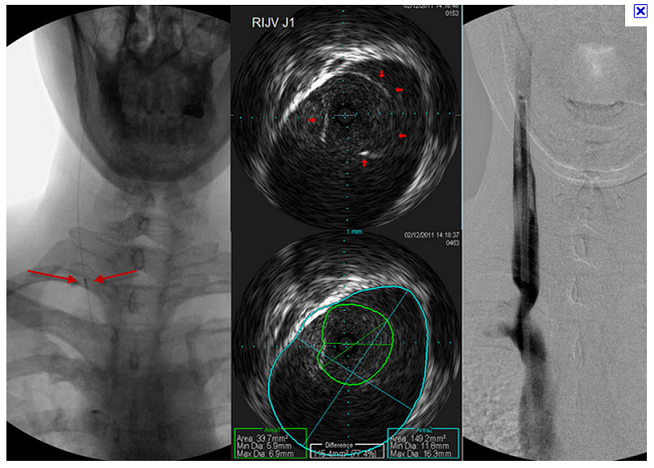
This is not the case of the week but a previous one. Ok, fluoroscopy only is the image on the left. Flouroscopy with the use of contrast is the image on the right. Both are in the jugular but only in the right one can the jugular be seen....

What does a balloon look like when it is not filled with contrast? Can waisting still be seen?
Last edited by Cece on Wed Jun 08, 2011 7:46 am, edited 1 time in total.
User avatar
pklittle
Family Elder
Posts: 341
Joined: Sun Mar 07, 2010 3:00 pm

Post by pklittle »

Dr Sclafani.. What an experience this woman has been going through! To be in a foreign country and have such a horrible reaction to the dye.

My question, as I usually feel, is so basic. How can you do the work with no liquid in the vein? How does IVUS work with no fluids? Aren't the veins limp and closed up everywhere whether there is stenosis or not?
User avatar
ozwannabe
Getting to Know You...
Posts: 16
Joined: Fri Dec 31, 2010 3:00 pm

Post by ozwannabe »

Hi Dr Sclafani,

I had venoplasty five weeks ago. I had complete occlusion of my LIJV and moderate RIJV. The left side had multiple collaterals involving vertebral plexus and the EJV. It was stenosed at the mandible and distal end. It was eventually dilated to 10.1mm. The right was stenosed at the distal end and was dilated to 12mm.

I have had lots of improvements and still getting stronger. Am wondering if these collaterals might affect venous drainage to the arm? The strangest thing is my watch is practically falling off its now so loose. Conversely my right hand has been going numb more than prior to the procedure. Could this be due to nerve irritation or restenoses? My RIJV initially spasmed and had worse flow but improved after additional balloonings.

I have concerns about my azygous as it was declared normal almost immediately after it was catheterised. It was surprisingly uncomfortable too compared to the stenosed veins. Will have to save up and visit New York to check it out properly.

Cheers,
Vicki
User avatar
drsclafani
Family Elder
Posts: 3182
Joined: Fri Mar 12, 2010 3:00 pm
Location: Brooklyn, New York
Contact:

Post by drsclafani »

Cece wrote:To use this fine example:
Image
This is not the case of the week but a previous one. Ok, fluoroscopy only is the image on the left. Flouroscopy with the use of contrast is the image on the right. Both are in the jugular but only in the right one can the jugular be seen....

What does a balloon look like when it is not filled with contrast? Can waisting still be seen?
fBallons are barely visible if at all without the use of contrast media. One can put air in it and you might see the balloon inflation as a black air like structure. In this case we put gadolinium in the balloon. its slight density is exploited
User avatar
drsclafani
Family Elder
Posts: 3182
Joined: Fri Mar 12, 2010 3:00 pm
Location: Brooklyn, New York
Contact:

Post by drsclafani »

pklittle wrote:Dr Sclafani.. What an experience this woman has been going through! To be in a foreign country and have such a horrible reaction to the dye.

My question, as I usually feel, is so basic. How can you do the work with no liquid in the vein? How does IVUS work with no fluids? Aren't the veins limp and closed up everywhere whether there is stenosis or not?
there is fluid in the vein. it is just not opacified. ivus does not require xray dye, it requires fluid.
the vfein has fluid and IVUS shows the problems.
User avatar
davmets2
Getting to Know You...
Posts: 21
Joined: Sun Nov 07, 2010 3:00 pm
Location: Albany, NY
Contact:

heat in back, chest discomfort & bladder retention probl

Post by davmets2 »

drsclafani,
I don't know if you remember my case. I had 2 procedures done back in January with Dr. Mehta. I came off of warifin on April 12th after 2 months. On the 12th the US said I had flow, but couldn't tell me how much. On May 6th I needed to go on solumedrol (3 days) with a 12 day taper. I seemed to have recovered ok from that for a couple weeks. Now all of a sudden, 2 weeks ago I seem to be having a major set back. I ended up in the ER, they tested my lungs (CT scan), was negative, heart seems to be fine. Bladder wasn't emptying, so they gave me a catheter for 5 days. I'm getting very worried that I'm in a very bad place that I may not recover from. I had an MRI of the spine and there were no active lesions to be seen. My neuro put me back on another 3 days of solumedrol last Friday with the taper again. Not feeling any better.
I'm really considering doing another procedure through you to see if this the real problem...I've got to do something. If you don't want to give me advice here, could you e-mail me with info to get in contact with you.
I've gone 33 years working and coping and all of a sudden I can't do either.
Thanks in advance.
Don
Cece
Family Elder
Posts: 9335
Joined: Mon Jan 04, 2010 3:00 pm
Contact:

Post by Cece »

Karen Moran at kmoran@aac-llc.com is the patient coordinator for making appts with Dr. Sclafani. It's ccsviliberation@gmail.com for his email.
drsclafani wrote:fBallons are barely visible if at all without the use of contrast media. One can put air in it and you might see the balloon inflation as a black air like structure. In this case we put gadolinium in the balloon. its slight density is exploited
glad there was a solution for that! Not seeing the balloons would've made things even more challenging.
Cece
Family Elder
Posts: 9335
Joined: Mon Jan 04, 2010 3:00 pm
Contact:

Re: case of the week

Post by Cece »

drsclafani wrote:The first image that struck me as unusual is #2. Here is a close-up:Image
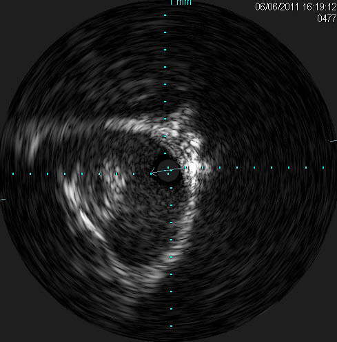
This is in the dural sinuses. The bright reflections do you see the tissue with reflections at about "9 o'clock?
i was wondering what this was. I have never seen anything like that before on IVUS. Could it be a clot?

I surmised that it represented Arachnoid granulations. These are SF-filled meningothelial-lined protrusions that are filled with CSF. They
extend into the venous sinuses through openings in the
dura. They filter CSF and drain it into central venous circulation.

it is important to differentiate them from dural sinus thrombosis and tumor. It was in a typical location for arachnoid granulations.
This is a very interesting case.
If it was an arachnoid granulation, nothing to treat there...what was the next image that struck you as unusual?
User avatar
drsclafani
Family Elder
Posts: 3182
Joined: Fri Mar 12, 2010 3:00 pm
Location: Brooklyn, New York
Contact:

Post by drsclafani »

ozwannabe wrote:Hi Dr Sclafani,

I had venoplasty five weeks ago. I had complete occlusion of my LIJV and moderate RIJV. The left side had multiple collaterals involving vertebral plexus and the EJV. It was stenosed at the mandible and distal end. It was eventually dilated to 10.1mm. The right was stenosed at the distal end and was dilated to 12mm.

I have had lots of improvements and still getting stronger. Am wondering if these collaterals might affect venous drainage to the arm? The strangest thing is my watch is practically falling off its now so loose. Conversely my right hand has been going numb more than prior to the procedure. Could this be due to nerve irritation or restenoses? My RIJV initially spasmed and had worse flow but improved after additional balloonings.

I have concerns about my azygous as it was declared normal almost immediately after it was catheterised. It was surprisingly uncomfortable too compared to the stenosed veins. Will have to save up and visit New York to check it out properly.

Cheers,
Vicki
vicki

only 12 mm? i truly hope that i am incorrect: I am going to guess that your symnptoms will return to baseline within four months. , maybe two. Dont quote me, tho..
User avatar
drsclafani
Family Elder
Posts: 3182
Joined: Fri Mar 12, 2010 3:00 pm
Location: Brooklyn, New York
Contact:

Re: case of the week

Post by drsclafani »

Cece wrote: If it was an arachnoid granulation, nothing to treat there...what was the next image that struck you as unusual?
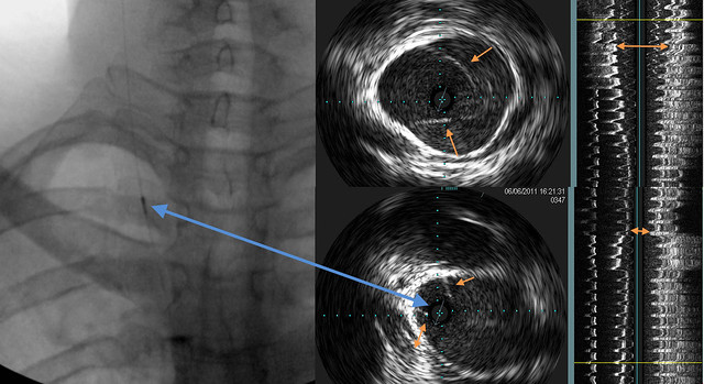
Let's review the VUS near the valve

Image

on the left is a fluoro picture of the IVUS with a blue arrow pointing to the IVUS device.
In the middle are two images of the valve. See how the blue arrow corresponds to the IVUS probe.follow the blue arrow to a small circle void of echos. If you look closely you will see a small arrow in the center of the echo-less circle. This represents the orientation of the images on the right. That image is a longitudinal view of the vein.

Going from left to right in the right image (got that?) there are several sites of reflections. These reflections represent the valve and the wall of the vein.
The orange arrows show progressive narrowing of the lumen of the vein.
User avatar
ozwannabe
Getting to Know You...
Posts: 16
Joined: Fri Dec 31, 2010 3:00 pm

Post by ozwannabe »

drsclafani wrote:
ozwannabe wrote:Hi Dr Sclafani,

I had venoplasty five weeks ago. I had complete occlusion of my LIJV and moderate RIJV. The left side had multiple collaterals involving vertebral plexus and the EJV. It was stenosed at the mandible and distal end. It was eventually dilated to 10.1mm. The right was stenosed at the distal end and was dilated to 12mm.

I have had lots of improvements and still getting stronger. Am wondering if these collaterals might affect venous drainage to the arm? The strangest thing is my watch is practically falling off its now so loose. Conversely my right hand has been going numb more than prior to the procedure. Could this be due to nerve irritation or restenoses? My RIJV initially spasmed and had worse flow but improved after additional balloonings.

I have concerns about my azygous as it was declared normal almost immediately after it was catheterised. It was surprisingly uncomfortable too compared to the stenosed veins. Will have to save up and visit New York to check it out properly.

Cheers,
Vicki
vicki

only 12 mm? i truly hope that i am incorrect: I am going to guess that your symnptoms will return to baseline within four months. , maybe two. Dont quote me, tho..
Thanks for your reply Dr Sclafani. Looks like maybe I better step up the savings plan.

Vicki
Cece
Family Elder
Posts: 9335
Joined: Mon Jan 04, 2010 3:00 pm
Contact:

Re: case of the week

Post by Cece »

drsclafani wrote:Let's review the VUS near the valve
Image

on the left is a fluoro picture of the IVUS with a blue arrow pointing to the IVUS device.
In the middle are two images of the valve. See how the blue arrow corresponds to the IVUS probe.follow the blue arrow to a small circle void of echos. If you look closely you will see a small arrow in the center of the echo-less circle. This represents the orientation of the images on the right. That image is a longitudinal view of the vein.

Going from left to right in the right image (got that?) there are several sites of reflections. These reflections represent the valve and the wall of the vein.
The orange arrows show progressive narrowing of the lumen of the vein.
ok, if we are standing on the tiny blue arrow, looking forward, we are seeing half of the vein longitudinally. If it is oriented this way, shouldn't the stenosis be on the right in the longitudinal image? But it looks like it is on the left.

In the longitudinal image, going from left to right, we are seing the vein wall, then the reflections off the valve, then the other vein wall, which is thicker?

The arrows very clearly show the narrowing.

In the IVUS images, are we seeing a healthy set of valves on the top image and then a second stumpy set of valves in the bottom image that is the stenosis? I originally thought the second image showed a continuation of the same valves in the first image.

And there is the 'bright bump' in the longitudinal image! The arrow showing the greatest narrowing is right at the start of the 'bright bump.' I had my perspective switched, I should've been looking at it as a narrowing, not as the widening below. But it is easier to see with the additional longitudinal image on top, if at the very top with the orange arrows is healthy vein. Always good to know what normal should be, when trying to identify abnormal....

Anyone else see what Dr. Sclafani is trying to show here?
User avatar
ozwannabe
Getting to Know You...
Posts: 16
Joined: Fri Dec 31, 2010 3:00 pm

Re: case of the week

Post by ozwannabe »

Cece wrote:
drsclafani wrote:Let's review the VUS near the valve

Image

on the left is a fluoro picture of the IVUS with a blue arrow pointing to the IVUS device.
In the middle are two images of the valve. See how the blue arrow corresponds to the IVUS probe.follow the blue arrow to a small circle void of echos. If you look closely you will see a small arrow in the center of the echo-less circle. This represents the orientation of the images on the right. That image is a longitudinal view of the vein.

Going from left to right in the right image (got that?) there are several sites of reflections. These reflections represent the valve and the wall of the vein.
The orange arrows show progressive narrowing of the lumen of the vein.
ok, if we are standing on the tiny blue arrow, looking forward, we are seeing half of the vein longitudinally. If it is oriented this way, shouldn't the stenosis be on the right in the longitudinal image? But it looks like it is on the left.

In the longitudinal image, going from left to right, we are seing the vein wall, then the reflections off the valve, then the other vein wall, which is thicker?

The arrows very clearly show the narrowing.

In the IVUS images, are we seeing a healthy set of valves on the top image and then a second stumpy set of valves in the bottom image that is the stenosis? I originally thought the second image showed a continuation of the same valves in the first image.

And there is the 'bright bump' in the longitudinal image! The arrow showing the greatest narrowing is right at the start of the 'bright bump.' I had my perspective switched, I should've been looking at it as a narrowing, not as the widening below. But it is easier to see with the additional longitudinal image on top, if at the very top with the orange arrows is healthy vein. Always good to know what normal should be, when trying to identify abnormal....

Anyone else see what Dr. Sclafani is trying to show here?
Hi , I'm taking a totally uneducated stab in the dark here. Is it a valve with fused leaflets on the narrow side?

Cheers,
Vicki
Cece
Family Elder
Posts: 9335
Joined: Mon Jan 04, 2010 3:00 pm
Contact:

Post by Cece »

Happy healthy restful vibes going out to Dr. Sclafani, who judging by his absence here is much too busy right now! How is the symposium coming along?
User avatar
drsclafani
Family Elder
Posts: 3182
Joined: Fri Mar 12, 2010 3:00 pm
Location: Brooklyn, New York
Contact:

Post by drsclafani »

Cece wrote:Happy healthy restful vibes going out to Dr. Sclafani, who judging by his absence here is much too busy right now! How is the symposium coming along?

help


the lectures are in preparation'
the rooms are secured
its a great program

But will they come?
registration is below projections.
Any help would be appreciated.

The link is www.ccsvisymposium.com

please spread the word to doctors and patient: learning about ccsvi
Post Reply
  • Similar Topics
    Replies
    Views
    Last post

Return to “Chronic Cerebrospinal Venous Insufficiency (CCSVI)”