Hello, I am a 39 year old male. I had an MRI done back in May for migraines. My primary Doctor adviced me to see a Neurologist because of some nonspecific spots. Which at the time didn't mean anything to me. Went to the Neurologist and he gave me some medications and in a few days my migraine was gone. Fast forward to a few weeks ago. I started getting some tingling feeling in my feet and hands, toes and fingers. Very intense at times. Also blurred vision. I went back to my regular doctor expecting him to tell me I was a diabetic and it was neuropathy. But my blood sugar was fine. He was very concerned and told me it more than likely had to do with the migraines and that I need to go back to the neurologist. Unfurtunately the town I live in is in short supply so my earliest appointment is Nov 28th. In the past week i have started having some other weird feeling (sensations). Balance off. Feel weak, like I have been working out. Left side of my face feels like I have been in a fight and lost! Pains shooting down my legs. Memory loss. And some other weird things. Anyways since i have to wait, i stated looking online. That was probably not the best idea. I got a copy of my MRI and was wondering if any of you good people good decipher it?
FINDING:
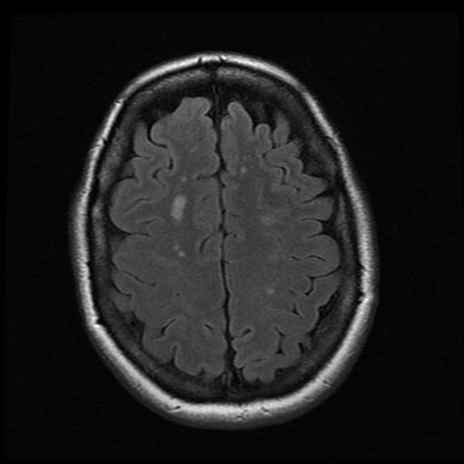
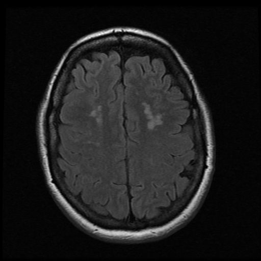
CEREBRUM: There are small foci of abnormal increased FLAIR signal in the subcortical white matter of both cerebral hemispheres. There is no MR evidence of mass, hemorrhage, or acute infarct.
CONCLUSION: Tiny foci of T2 hyperintensity in the subcortical white matter are nonspecific. Differential diagnosis includes microvascular ischemic changes, demyelinating disorders, and dysmyelinating disorders.
Any help would be greatly appreciated
MRI Report?
-
cheerleader

- Family Elder
- Posts: 5361
- Joined: Mon Sep 10, 2007 2:00 pm
- Location: southern California
Re: MRI Report?
Hey djham---
Migraine often shows these kind of non-specific white matter changes. Sorry you've found yourself here! Be careful reading too much stuff on MS online...the mind can do lots of stuff to the body. An MS diagnosis is made after going thru about 100 differential diagnoses, and migraine and ischemic (low oxygen) changes are on the list. That's why your MRI says "non specific"...that's good news. MS lesions are often found in the corpus callosum area of the brain, or in the deep periventricular white matter, or look like Dawson's Fingers, and have an "ovoid" shape, (perpendicular to the long axis of the lateral ventricles.) Or be found on the cervical spine. I learned this because my husband's lesions were in the corpus callosum, and on the spine...indicative of MS. The fact yours are tiny foci is good news, too. The odds are it's probably not MS.
While you're waiting for more specific testing for MS, like another MRI with contrast dye, or a lumbar puncture---take care of yourself. Eat a lower fat diet, high in antioxidants with lots of fruits and veggies. Quit smoking, limit alcohol. Look into a vitamin D supplement, cut out sweets, try to reduce stress, get good sleep, and make sure to exercise. All of these measures will help your general health, and are shown to help oxidative stress--something in MS and all neurodegenerative brain issues. And these measures would help if you have small vessel disease of the brain and cardiovascular issues---much more likely to be your situation.
Here's more info on MRIS for you---
When MRIs are ordered, 50% of people show white matter lesions, and these are almost always vascular in nature.
Hope this info helps. Be well,
cheer
Migraine often shows these kind of non-specific white matter changes. Sorry you've found yourself here! Be careful reading too much stuff on MS online...the mind can do lots of stuff to the body. An MS diagnosis is made after going thru about 100 differential diagnoses, and migraine and ischemic (low oxygen) changes are on the list. That's why your MRI says "non specific"...that's good news. MS lesions are often found in the corpus callosum area of the brain, or in the deep periventricular white matter, or look like Dawson's Fingers, and have an "ovoid" shape, (perpendicular to the long axis of the lateral ventricles.) Or be found on the cervical spine. I learned this because my husband's lesions were in the corpus callosum, and on the spine...indicative of MS. The fact yours are tiny foci is good news, too. The odds are it's probably not MS.
While you're waiting for more specific testing for MS, like another MRI with contrast dye, or a lumbar puncture---take care of yourself. Eat a lower fat diet, high in antioxidants with lots of fruits and veggies. Quit smoking, limit alcohol. Look into a vitamin D supplement, cut out sweets, try to reduce stress, get good sleep, and make sure to exercise. All of these measures will help your general health, and are shown to help oxidative stress--something in MS and all neurodegenerative brain issues. And these measures would help if you have small vessel disease of the brain and cardiovascular issues---much more likely to be your situation.
http://braindiseases.wordpress.com/2008 ... e-answers/A lot of work has been done to determine the significance of white matter lesions. The thinking now is that they represent ischemia (lack of blood flow) in the small blood vessels of the brain. Hence they are also at times referred to as ischemic small vessel disease. Hence these lesions are more commonly seen in the MRI of patients who have cerebrovascular risk factors like hypertension, diabetes and high cholesterol as well those that smoke. Their incidence increases as we age (meaning you are more likely to see them on the MRI of someone who is 60 and above rather than someone who is in his 20′s).
Here's more info on MRIS for you---
When MRIs are ordered, 50% of people show white matter lesions, and these are almost always vascular in nature.
http://www.radiologyassistant.nl/en/4556dea65db62Consequently, it is not wise to put MS in the differential diagnosis, if the clinician does not suspect the patient of having MS and on the MR incidental WMLs are found.
The odds are against the diagnosis of MS, because vascular WMLs are 50-500 times more likely than MS plaques.
On the other hand if a patient is clinically suspected of having MS and multiple WMLs are found, our major concern is the differential diagnosis MS versus vascular disease and we have to follow the McDonald criteria.
Hope this info helps. Be well,
cheer
Husband dx RRMS 3/07
dx dual jugular vein stenosis (CCSVI) 4/09
http://ccsviinms.blogspot.com
dx dual jugular vein stenosis (CCSVI) 4/09
http://ccsviinms.blogspot.com
-
misticrowder
- Newbie
- Posts: 1
- Joined: Tue Aug 20, 2013 5:13 am
Re: MRI Report?
Hello, I have been experiencing some of the same problems but also one side of my face and tongue going numb and weird vibrating sensations. Im being tested for MS and my MRI showed tiny foci as well. Did you ever get a diagnosis?? Please let me know thanks!
-
centenarian100
- Family Elder
- Posts: 504
- Joined: Mon Apr 15, 2013 9:51 am
Re: MRI Report?
Hey djham.
A few questions:
Can you describe your blurry vision? Is it in one eye or both? Do you have difficulty seeing in a specific area of your visual field (scotoma)? Do you have pain with eye movement? Cover each eye and look at something red-do you note a difference in the intensity of the red color in one eye (red color desaturation)?
Can you describe your other symptoms in greater detail?
Do you happen to have the CDs for your MRI? If you could upload a few of the axial T2 FLAIR images showing the lesions, that might be helpful.
The radiologist hedges in the read, so the text itself isn't very helpful. Often, imaging in MS is non-specific and must be interpreted in the appropriate clinical context. Even well demarcated callosal lesions can occur in other neurological conditions (i.e. Sussac syndrome).
A few radiology jokes for you:
What is the radiologist's favorite plant? The hedge
What is the radiologist's favorite breakfast food? The waffle
Don't stress yourself out too much. Uncertainty can be more psychologically damaging than outright bad news. Best of luck to you. Definitely take cheerleader's advice on general healthy living.
-c
-
gunzgirl07
- Newbie
- Posts: 2
- Joined: Thu Jan 08, 2015 11:18 am
MRI not sure
So I went to my go with fatigue, headaches memory issues, pins and needles in legs, fogginess. Sent me for a mri results came back with: best seen on FLAIR several areas of increased signal intensity biaterally in predominantly subcritical white matter. Impression: demyelinating disease or infectious etiology including Lyme's. Have no idea what any of this means I went for blood wrk n show no trace for Lymes. Not sure what this means
-
lyndacarol

- Family Elder
- Posts: 3394
- Joined: Thu Dec 22, 2005 3:00 pm
- Contact:
Re: MRI not sure
Welcome to ThisIsMS, gunzgirl07.gunzgirl07 wrote:So I went to my go with fatigue, headaches memory issues, pins and needles in legs, fogginess. Sent me for a mri results came back with: best seen on FLAIR several areas of increased signal intensity biaterally in predominantly subcritical white matter. Impression: demyelinating disease or infectious etiology including Lyme's. Have no idea what any of this means I went for blood wrk n show no trace for Lymes. Not sure what this means
I have no medical background, but, in my opinion, when you presented to your GP with "fatigue, headaches, memory issues, pins and needles in legs, fogginess" (which are classic and consistent with vitamin B12 deficiency), the first condition your GP should have considered and ruled out is a possible B12 deficiency by ordering a blood test: the HoloTranscobalamin (HoloTc) test. (The serum B12 test alone is unreliable and inadequate for determining a deficiency.)
Request a copy of any test results you have done. You need to have the actual numbers!
By the way, neurological lesions can be a sign of B12 deficiency as well as a sign of demyelinating disease, such as MS. Both B12 deficiency and MS affect the myelin; you can't tell them apart, except by testing.
-
gunzgirl07
- Newbie
- Posts: 2
- Joined: Thu Jan 08, 2015 11:18 am
Re: MRI Report?
Thank you!!! Go didn't send for b12 blood work. I was sent for Lyme's n others. I saw a neuro and she stated that MRI is nothing to wrry about bc of my age (32) that all it is white spots that can b bc I quit smoking (3 yrs ago). She stated that the pins and needles are from sciatic which yes I have disc issues for 10 yrs but never had pins and needles like I can stop moving them. I don't want it to b serious but neuro doesn't care to listen to fogginess, memory issues headaches r from stress.
-
callmestitch
- Newbie
- Posts: 4
- Joined: Mon Jan 12, 2015 4:44 pm
Re: MRI Report?
Hi all,
wondering what you all think, I have pretty much same sx as stated in these posts, also had slurred speech, facial palsy, sore eye movement in one eye, pins n needles on one side in toes then graduating to whole foot, bad cognition and word finding, had MRI done and report was as follows:
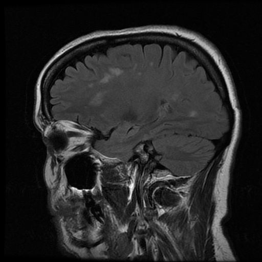
Best appreciated on the FLAIR sequence, there is periventricular high signal seen together with scattered foci of high signal within the deep white matter seen in a pericallosal distribution. This is more pronounced than what would be expected for a patient of stated age of 43 years.
Best appreciated on the sagittal FLAIR, there are multiple T2 hyperintensities seen at the callosal septal interface with a perpendicular orientation. These do not demonstrate significant enhancement. The main differential for this would be that of demyelination. Progress imaging is recommended.
have included some images, my neuro says possible M.S right off the bat, but wants to see what my latest MRI (last week) shows, am yet to get report from that one or images.
do you think this could be M.S?
not sure what it all means though
cheers
Stitch
wondering what you all think, I have pretty much same sx as stated in these posts, also had slurred speech, facial palsy, sore eye movement in one eye, pins n needles on one side in toes then graduating to whole foot, bad cognition and word finding, had MRI done and report was as follows:
Best appreciated on the FLAIR sequence, there is periventricular high signal seen together with scattered foci of high signal within the deep white matter seen in a pericallosal distribution. This is more pronounced than what would be expected for a patient of stated age of 43 years.
Best appreciated on the sagittal FLAIR, there are multiple T2 hyperintensities seen at the callosal septal interface with a perpendicular orientation. These do not demonstrate significant enhancement. The main differential for this would be that of demyelination. Progress imaging is recommended.
have included some images, my neuro says possible M.S right off the bat, but wants to see what my latest MRI (last week) shows, am yet to get report from that one or images.
do you think this could be M.S?
not sure what it all means though
cheers
Stitch
-
lyndacarol
- Family Elder
- Posts: 3394
- Joined: Thu Dec 22, 2005 3:00 pm
- Contact:
Re: MRI Report?
Hi and welcome to ThisIsMS, Stitch (callmestitch).
I have no medical background, but to answer your question… yes, based on your images and description (especially the periventricular location), I think it could be MS. But MS is a diagnosis of exclusion, made after other more likely possible causes have been ruled out.
Since all your symptoms (even neurological lesions) are consistent with vitamin B12 deficiency, it is my opinion that your neuro should rule out this possibility first, with a HoloTranscobalamin (HoloTc) blood test. This newer test, which tests for B12 on the transcobalamin II (the only transporter protein that can carry the vitamin into the tissues/cells), is considered to be much more accurate in determining a deficiency than the older serum B12 test, which measures total B12 (80% of which can be bound to another protein incapable of getting B12 into the cells).
It is my opinion that this is the place to start.
I urge you to read the book, Could It Be B12? An Epidemic of Misdiagnoses by Sally M. Pacholok, RN, BSN, and Jeffrey J. Stuart, D.O. (This may be available at your library.): http://b12awareness.org/could-it-be-b12 ... diagnoses/
-
callmestitch
- Newbie
- Posts: 4
- Joined: Mon Jan 12, 2015 4:44 pm
Re: MRI Report?
Hi lindacarol
sorry if my pictures turned out too big, am new at posting pictures, will haveto shrink them a little so they don't take up sooo much room lol oops
I forgot to mention, My Neuro is pretty good, did all manner of blood tests including B12 which was in normal range. Also forgot to mention I was hyperthyroid and had the radioactive iodine treatment which sent me to hypothyroid, for some time but my levels are coming back to normal now, we found some of my symptoms correlated to this but not all. The lesions on brain were discovered routinely as they always do an MRI on people with facial palsy and slurred speech as a precaution as it might have been a small stroke (it wasn't)
I am not enjoying the dx process though, the limbo you tend to end up in (ie could be, might be, is possibly) can be a little frustrating, that MRI was taken a year ago, and my sx are off and on now, not as severe. A new sx was sudden onset of Migraines, had 3 in as many consecutive days, which had my Neuro a little concerned as I have no history of migraine let alone bad headaches. Would M.S cause Migraine also? I have not heard of it as a symptom, but have read some things online where majority of M.Sers have migraines, but you never can trust everything you read hey.. lol
Thanks for your reply
be well
Stitch
-
lyndacarol
- Family Elder
- Posts: 3394
- Joined: Thu Dec 22, 2005 3:00 pm
- Contact:
Re: MRI Report?
I agree with you, Stitch, that your neuro, who "did all manner of blood tests including B12," sounds very good. (If available, are you willing to share the number results for the HoloTc test? – You may understandably consider this too personal and not wish to share.) He seems to be well on his way to ruling out possibilities: B12 deficiency, thyroid problems, etc.callmestitch wrote:My Neuro is pretty good, did all manner of blood tests including B12 which was in normal range. Also forgot to mention I was hyperthyroid and had the radioactive iodine treatment which sent me to hypothyroid, for some time but my levels are coming back to normal now, we found some of my symptoms correlated to this but not all. The lesions on brain were discovered routinely as they always do an MRI on people with facial palsy and slurred speech as a precaution as it might have been a small stroke (it wasn't)
I am not enjoying the dx process though, the limbo you tend to end up in (ie could be, might be, is possibly) can be a little frustrating, that MRI was taken a year ago, and my sx are off and on now, not as severe. A new sx was sudden onset of Migraines, had 3 in as many consecutive days, which had my Neuro a little concerned as I have no history of migraine let alone bad headaches. Would M.S cause Migraine also? I have not heard of it as a symptom, but have read some things online where majority of M.Sers have migraines, but you never can trust everything you read hey.. lol
Your symptom of "pins n needles on one side of the toes then graduating to whole foot" is the textbook definition of "peripheral neuropathy." This is a common symptom in many conditions. In investigating the cause of peripheral neuropathy, the University of Chicago suggests the following:
http://peripheralneuropathycenter.uchic ... #bloodtest\
To your question, "Would MS cause migraine?"… Once again, migraine headaches are common to many conditions, including among those listed above, celiac disease/gluten sensitivity. Has your Dr. screened for this and ruled it out? Has he considered other possibilities on this list?Blood tests
Blood tests are commonly employed to check for vitamin deficiencies, toxic elements and evidence of an abnormal immune response.
Depending on your individual situation, your doctor may request certain laboratory tests to identify potentially treatable causes for neuropathy. These include tests for:
Vitamin B12 and folate levels
Thyroid, liver and kidney functions
Vasculitis evaluation
Oral glucose tolerance test
Antibodies to nerve components (e.g., anti-MAG antibody)
Antibodies related to celiac disease
Lyme disease
HIV/AIDS
Hepatitis C and B
The limbo of the diagnostic process is absolutely frustrating. Unfortunately, there is no definitive test for MS – it is a case of "last man standing" after the others have been ruled out.
We wish you all the best, and are here to answer any questions that we can.
Last edited by lyndacarol on Tue Jan 13, 2015 3:49 pm, edited 1 time in total.
-
callmestitch
- Newbie
- Posts: 4
- Joined: Mon Jan 12, 2015 4:44 pm
Re: MRI Report?
lyndacarol,
unfortunately, I don't have alot of the test results, they only gave me the disk of the MRI, alot of test results were sent to my regular GP just to keep him updated (he's a really good GP I've had for years will even call you up to see how you are) I tend to lose alot of paperwork so am happy to keep my results with him and he is happy to keep my file if ever I should need it. I forget where I put things then accidently throw stuff out. I know when I was in the hospital (inital attack put me in hospital for 8 days, my Neuro had me tested for countless things and was very through, she did say she ruled out the things that mimic, but she is currently looking or questioning if it is also small vein something or other in the brain that is causing the lesions, not entirely sure what she called it, but she did say they haveto be sure to look at it if not to rule it out also. Other than the symptoms I've had I'm basically healthy and 'normal' was what she stated. I did have problems with some reflexes, but they seemed to fix themselves. Though I have experienced cramping on foot and toes and calf muscle slightly. But these things I will bring up with her next time I see her. My partner is worried it could be M.S but I have done some reading on it and it does not have me too worried, not like it is a death sentence hey.
Thanks for your reply again
be well
Stitch
-
callmestitch
- Newbie
- Posts: 4
- Joined: Mon Jan 12, 2015 4:44 pm
Re: MRI Report?
Hi again,
just a couple of questions..
I've seen people who post their scans that show one lesion on the brain and they get the dx of M.S, why wouldn't a Dr diagnose M.S with the amount I have, just because there is no change? they have ruled out everything else that mimic's M.S, and say it is unusual for someone my age to have these lesions (I'm 45yrs old) My latest MRI shows no change but I have read that a lesion may not show an enhancement with contrast because a flair might be over. Just because a lesion doesnt enhance when the dr views results does that mean you do not have MS or the timing was out? How big do dawsons fingers haveto be? I know my neuro states possible M.S, and some sx had nothing to do with my thyroid at the time (leading her to state possible M.S) but why come out and state it if it is not. very frustrating, and not only that, why can't they give reason to such lesions if it is not M.S, feel like I've been left hanging wondering well.. what is it then, what caused these lesions etc
feeling frustrated
Stitch
-
harvinder1988
- Newbie
- Posts: 1
- Joined: Sun Nov 12, 2017 9:41 pm
Re: MRI Report?
hi, m 30 yrs old feeling tingling in my hands & legs and some weakness in my left hand and left eye twitching often.Also having pain in my neck and left shoulder going to my left hand.
I have done MRI of my brain & C-Spine,findings are below:-
MRI - C spine
Sequences- T1w & T2w, Axial - T1w & T2w
Impressions:-
The study reveals straightening of normal cervical spine
There is diffuse bulge at C3-C4 level intending the thecal sac.
MRI Brain
Performed on high resolution T1-Se,T2-FSE,T2-Flair,GRE Axial,T-2 Flair,T2-FSE Coronal,T2-FSE,T1-SE saggital diffusion weighted using head coil on 1.5 Tesla scanner.
Impressions:-
there are evidence of few foci of altered signal intensity appearing hyperintense on T2 and Flair sequence seen involving the bilateral periventricular white matter and centrum semivale.the findings are suggestive of non specific white matter lesion.
There is evidence of Mucosal thickening seen involving the floor of right maxillary sinus.
What it means?
Can i suffering from MS?
Re: MRI Report?
Hi harvinder1988,
The best thing you can do is consult with a Dr. as to the meaning of your MRIs and reports.
My unprofessional interpretation:
Bulging and herniated discs can cause Neurological symptoms but this is not due to a disease such as MS. A Neurosurgeon/Orthopedist should be consulted. Back problems are sometimes helped with Physical Therapy but sometimes surgery is needed.
From your Brain MRI:
The best thing you can do is consult with a Dr. as to the meaning of your MRIs and reports.
My unprofessional interpretation:
.There is diffuse bulge at C3-C4 level intending the thecal sac
Bulging and herniated discs can cause Neurological symptoms but this is not due to a disease such as MS. A Neurosurgeon/Orthopedist should be consulted. Back problems are sometimes helped with Physical Therapy but sometimes surgery is needed.
From your Brain MRI:
Non-specific lesion means just that. Brain lesions can be due to just about anything and doesn't mean a person has MS.findings are suggestive of non specific white matter lesion