Hi everyone,
I have recently had the procedure done by Dr Ludgya (and Simka) in Poland.
I had tried to get the UK dr's to come on board (which now they seem to be) but at the time they would not agree to do a venogram.
My MRV and CT came back normal as well as the doppler verified by 2 UK Vascular specialists. (they have now seen the confirmation of a problem on the Venogram and I believe that is why they are coming on board)
I then sent my scans to Dr Dake. He saw that both my jugular veins were pancaked flat at C2 and a further occlusion was seen in the lower left jugular
I went to Poland to be treated by Dr Simka and he saw the problem with the left lower jugular it was obvious but did not see the stenosis that Dr Dake had seen high up. I did mention it but he didn't think it was relevant or too risky to treat (not sure)
I had the venography which confirmed the lower left jugular occlusion and this was treated by ballooning. I had lots of collaterals right up high by the jaw line.
My worry is that because the high stenosis seen by Dr Dake was not treated that my problem may not be truly solved.
I might send my Venogram to Dr Dake to ask his opinion in this.
Any thoughts?
(I have had some improvements: eyes, energy and not feeling MSy but have had a couple of bad days with weak arms, that seems to be easing off now which could be down to CCSVI but I think it is down to regimen change)
Different approach to treating CCSVI Dake and Simka
-
LR1234
- Family Elder
- Posts: 1517
- Joined: Wed Feb 11, 2009 3:00 pm
- Location: California
- Contact:
Different approach to treating CCSVI Dake and Simka
Last edited by LR1234 on Sun Jan 31, 2010 3:26 am, edited 3 times in total.
-
wonky1

- Family Elder
- Posts: 113
- Joined: Thu Oct 08, 2009 2:00 pm
- Contact:
Hi LR
I had a similar concern. I had stenosis high up in both jugs but he felt this was a secondary problem and resulted from the blockages further down the pipe. The stenosis was actually just a collapsed vein due to there being no flow to keep it open.
I am pasting below the email he sent in response to my question.
This is really the open question. At Stanford such a narrowing is regarded as real problem, and they usually put a stent in this area. But our interpretation is that it is only a collapse of the vein, which is secondary to diminished flow caused by problems in the lower part of the vein. This does not apply to all patients, but in your case our interpretation was as steted above, since the upper stenosis was not visible after infection of dye (during the procedure) and also in was not visible after ballooning the stenosis. However, narrowing of both jugular veins was still visible using Doppler on the day after operation, but we usually see that the improvement in flow rates takes more than one or two days. We will see what happens to these narrowings during your second visit to Poland.
Happy New Year
Marian Simka
[/i]
-
LR1234
- Family Elder
- Posts: 1517
- Joined: Wed Feb 11, 2009 3:00 pm
- Location: California
- Contact:
So Wonky, have you gone back to Poland? and did they look for you at these high stenosis? Also did they check your vertebral veins (I would also feel more comfortable having these checked).
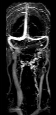
I have asked Mark, to post a couple of scans that I have pulled out of the video to show what I mean. In the first scan (Pre-op) the dye is placed very high and all the collaterals are sooo obvious. Then the second scan (post-op) the collaterals look like they have gone but I have realised that the dye was placed lower so maybe if the dye was put in higher again the collaterals will still be there. Also I had no treatment on my right jugular.
Maybe I need to look at those scans again of the right jugular at C2 to see if its flowing ok.
-
wonky1
- Family Elder
- Posts: 113
- Joined: Thu Oct 08, 2009 2:00 pm
- Contact:
Hi LR
Yes I just returned, see the thread Poland part 2.
We didn't discuss the vertebral veins. I think they saw they were clear from the MRI during the first visit.
I didn't discuss the high stenosis either. I surmise from the fact it was not raised that he was correct in his email that the stenosis was secondary to the valve problem and opened again when flow was restored.
-
LR1234
- Family Elder
- Posts: 1517
- Joined: Wed Feb 11, 2009 3:00 pm
- Location: California
- Contact:
-
cheerleader

- Family Elder
- Posts: 5361
- Joined: Mon Sep 10, 2007 2:00 pm
- Location: southern California
These are separate problems. I would not characterize the doctors as using different approaches. Each MS patient will be different. Just because one shows high stenosis with lower occlusion, does not mean another can be treated in the same method or has the same valve issues. I think we risk getting into trouble second guessing vascular and interventional radiologists, or playing doctor.
In my Jeff's case, the lower jugulars were open just fine. Valves looked great. This is why the neck doppler test showed no problem. It was the pinching of the high jugular that created the collaterals, which disappeared after stenting, and have remained gone. Dr. Dake spent a lot of hours in the vertebral veins, azygos and lower jugulars twice...two four hour procedures-scanning the flow at all the different junctions. The venogram is the true test. We are confidant in his diagnosis and treatment, and have seen evidence on the correction in the monitoring of flow. He is our local doctor, and we are able to contact him for follow up and questions.
This has been my concern for patients traveling thousands of miles to receive treatment, and why I continue to urge a relationship with local doctors for followup. I know it's been tough for you to find anyone in the UK, L...and I completely sympathize. Sorry to seem brusque, but this distance issue is a concern for followup.
cheer
Husband dx RRMS 3/07
dx dual jugular vein stenosis (CCSVI) 4/09
http://ccsviinms.blogspot.com
dx dual jugular vein stenosis (CCSVI) 4/09
http://ccsviinms.blogspot.com
-
LR1234
- Family Elder
- Posts: 1517
- Joined: Wed Feb 11, 2009 3:00 pm
- Location: California
- Contact:
Thanks Cheer,
The thing is other than that high stenosis nothing was done to the right jugular as I had no lower issues on the right. (maybe like Jeff)
I suppose as more research is done the anwser will become apparant and I will be treated again if need be:)
I just want to make sure everything is flowing as well as it can.
Last edited by LR1234 on Sun Jan 31, 2010 2:28 pm, edited 1 time in total.
-
CureIous

- Family Elder
- Posts: 1266
- Joined: Tue Jul 14, 2009 2:00 pm
- Location: Riverside, CA
- Contact:
LR's scans
Posting these for LR1234.
BEFORE

AFTER

Note the various collaterals that vanished on the after.
G'Day!
Mark
BEFORE
AFTER
Note the various collaterals that vanished on the after.
G'Day!
Mark
RRMS Dx'd 2007, first episode 2004. Bilateral stent placement, 3 on left, 1 stent on right, at Stanford August 2009. Watch my operation video: http://www.youtube.com/watch?v=cwc6QlLVtko, Virtually symptom free since, no relap
-
LR1234
- Family Elder
- Posts: 1517
- Joined: Wed Feb 11, 2009 3:00 pm
- Location: California
- Contact:
-
shye
- Family Elder
- Posts: 758
- Joined: Sun Nov 29, 2009 3:00 pm
- Location: NYC
- Contact:
-
cheerleader
- Family Elder
- Posts: 5361
- Joined: Mon Sep 10, 2007 2:00 pm
- Location: southern California
Just for comparison, you can see how L's collaterals were lower and continued into her lower neck and chest. Jeff's were very high, but the lower jugular is open. Again, I do not believe Dake and Simka are treating any differently, I believe that what my husband presented with was very different than L, and treated correctly for his case. Each MS patient is unique.
Husband dx RRMS 3/07
dx dual jugular vein stenosis (CCSVI) 4/09
http://ccsviinms.blogspot.com
dx dual jugular vein stenosis (CCSVI) 4/09
http://ccsviinms.blogspot.com
-
patientx
- Family Elder
- Posts: 1072
- Joined: Wed Sep 10, 2008 2:00 pm
LR:
You might want to read the second to last post here:
http://www.thisisms.com/ftopic-9905-15.html
You might want to read the second to last post here:
http://www.thisisms.com/ftopic-9905-15.html
-
cheerleader
- Family Elder
- Posts: 5361
- Joined: Mon Sep 10, 2007 2:00 pm
- Location: southern California
The best thing to do is to consult a doctor. We are only patients and caregivers on this site, and playing guessing games about where stenosis is located is not a good idea. I know some folks have their "theories" about this, but they are not privy to any outside information. Dr. Zamboni and Dr. Dake have spoken at length about this. Jeff has another location of stenosis very high above the jugular that cannot be stented, but it has been ballooned. We hope for the best. There are other patients showing up with this very high stenosis (into the sinus) In CCSVI stenosis can be located in any place outside the cranium, and only with venography can the exact location be found. period.
cheer
cheer
Husband dx RRMS 3/07
dx dual jugular vein stenosis (CCSVI) 4/09
http://ccsviinms.blogspot.com
dx dual jugular vein stenosis (CCSVI) 4/09
http://ccsviinms.blogspot.com
-
Arcee
- Family Elder
- Posts: 338
- Joined: Wed Jan 05, 2005 3:00 pm
- Location: Massachusetts, USA
- Contact:
And another thing to consider is that Dr. Dake and Dr. Simka are perfectly capable of having direct conversations about this kind of thing and seeing if there are, in fact, differences in how they approach things. They very well may have done so, and maybe they will (again) at the upcoming conference.
diagnosed RR in spring '04
1 stent placed in left jugular vein 7/15/09
on and off Copaxone
allergric to interferons and Tysabri
1 stent placed in left jugular vein 7/15/09
on and off Copaxone
allergric to interferons and Tysabri
-
patientx
- Family Elder
- Posts: 1072
- Joined: Wed Sep 10, 2008 2:00 pm
No one suggested otherwise. Although it seems people don't mind playing amateur radiologist.The best thing to do is to consult a doctor....
LR had concerns about a difference of opinion in her case. It seems others have run into the same concerns, so it might be useful to hear what they have to say, regarding the opinions of other vascular experts. I don't know what these "theories" are, but not all doctors agree on all aspects of CCSVI - including, it appears, Dr. Simka himself.