drsclafani wrote:Yes Cece
although you have a copy of the images, perhaps it would be good for you and your fans to review here as you suggest.
The sequence was as follows
1. prep and drape
2. time out
3. cath in left femoral under local anesthesia
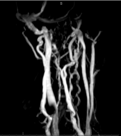
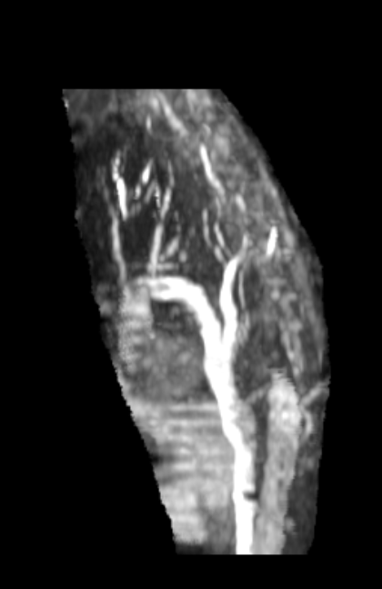
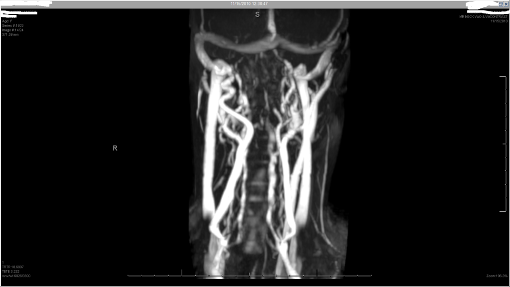
4. advancement into right jugular, venogram, ivus, 18 mm angioplasty to thirteen atmospheres, repeat venogram
5. advancement into left jugular, venogram, ivus, angiopasty to 14 mm at ten Atm, repeat venogram
6. advancement into azygous vein, venogram of ascending azygous followed by zero, twenty, seventy and ninety degree views of the azygous arch
7. IVC venogram
8 ascending lumbar venogram
13 atm and 10 atm! Those are not high pressures.
images as follows
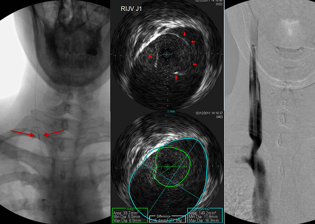
This sequence shows the ivus probe on the far left of the screen at the point where these images were taken. The venogram is on the right shows a bad stenosis.
look at the ivus probe! so small.
I didn't know you could compare the two area measurements so easily like that.
My goal has become to stretch the narrowing as much as possible without stretching the area of normal vein above it. That is a great value of the IVUS.
Everything he mentions here, about sparing the vein above the stenosis, and determining that the upper narrowing is phasic and not stenotic, the goal is less damage to our veins.
I've heard the story now, from bestadmom's perspective and from Dr. Sclafani's perspective, of how he was intrigued into CCSVI, but I marvel at how we as a group and I personally have now benefited from his taking an interest in this.
I am in danger of rhapsodizing again, I'd better move on....
more later....


 [/img]