Cece wrote:drsclafani wrote:So first i looked at the left renal vein:
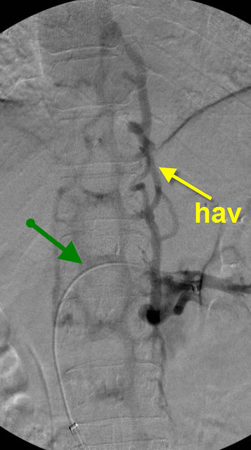
The green arrow points to the catheter in the renal vein. There is no contrast in the renal vein. rather the renal vein drains through the hemiazygous vein and outlines all the lumbar veins and the vertebral plexus.
This is a tremendous amount of blood to add to the azygous vein and vertebral plexus. The left renal vein drains about 500 ml of blood/minute. The entire brain drains approximately 750 ml of blood/minute. Thus this venous obstruction almost doubles the blood in the cerebrospinal blood pool, not good when the aygous and its collaterals, and the the internal jugular veins have stenoses!
500 ml/minute!
I am a bit aghast, I did not know the renal vein drained that much. Previously you had said you'd found one case of renal stenosis, now it presumably is just two cases out of 150. But when found, it is significant.
I can check google scholar for this myself but it's a question raised: what is typically done for renal vein stenosis? Is this an uncommonly treated area or is there a body of research on it?
I know there is controversy in whether to stent or not stent the iliac vein. I have heard very little about the renal vein.
Thanks for asking
yes i have now two cases. both have azygous disease.
The first patient had very very short lived clinical improvements before things got worse.
I did not treat the renal vein because i thought that i needed to do further workup before i addressed it.
the usual workup includes a urinalysis for red blood cells, protein. checks of blood pressure which may be elevated. queries regarding back pain, and pelvic pain.
Computed tomography is recommended for futher evaluation of the number of veins, and their locations.
the usual cauases of this type of obstruction is compression due to adjacent tumors and lymph nodes, compression between the aorta and the superior mesenteric artery (NUTCRACKER SYNDROME), or aberrant location of the renal vein behind the aorta (RETROAORTIC RENAL VEIN) or the vein splits into two with one going behind and one going in front of the aorta (CIRCUMAORTIC RENAL COLLAR).
Treatments include surgical disentanglement by moving these major veins and resuturing in a way that takes the pressure off, or nephrectomy, or stenting. Stenting has come into favor because it is so much less invasive.
another cause is thrombosis of the renal vein. This is much less likely to occur
i think that these are truncal malformations in many circumstances wheree the veins of the fetus malform during transition between the fetal veins and the adult veins.