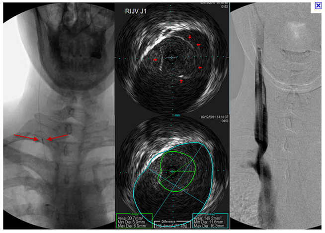
drsclafani wrote:Let's review the VUS near the valve
on the left is a fluoro picture of the IVUS with a blue arrow pointing to the IVUS device.
In the middle are two images of the valve. See how the blue arrow corresponds to the IVUS probe.follow the blue arrow to a small circle void of echos. If you look closely you will see a small arrow in the center of the echo-less circle. This represents the orientation of the images on the right. That image is a longitudinal view of the vein.
Going from left to right in the right image (got that?) there are several sites of reflections. These reflections represent the valve and the wall of the vein.
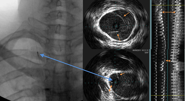
The orange arrows show progressive narrowing of the lumen of the vein.
ok, if we are standing on the tiny blue arrow, looking forward, we are seeing half of the vein longitudinally. If it is oriented this way, shouldn't the stenosis be on the right in the longitudinal image? But it looks like it is on the left.
In the longitudinal image, going from left to right, we are seing the vein wall, then the reflections off the valve, then the other vein wall, which is thicker?
The arrows very clearly show the narrowing.
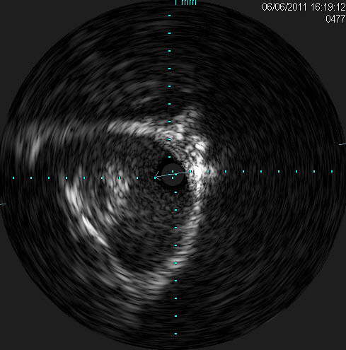
In the IVUS images, are we seeing a healthy set of valves on the top image and then a second stumpy set of valves in the bottom image that is the stenosis? I originally thought the second image showed a continuation of the same valves in the first image.
And there is the 'bright bump' in the longitudinal image! The arrow showing the greatest narrowing is right at the start of the 'bright bump.' I had my perspective switched, I should've been looking at it as a narrowing, not as the widening below. But it is easier to see with the additional longitudinal image on top, if at the very top with the orange arrows is healthy vein. Always good to know what normal should be, when trying to identify abnormal....
Anyone else see what Dr. Sclafani is trying to show here?