
The back flow down the right Iliac vein is due to the compression from the right Iliac artery as I mentioned above. Your heart is just fine. Now you need a stent on the right Iliac vein too.
Make sure you show your doctor the picture I have posted above.
Good luck
May Thurner. Just what are the symptoms?
-
AlmostClever
- Family Elder
- Posts: 366
- Joined: Mon Dec 21, 2009 3:00 pm
- Location: Houston, TX
- Contact:
-
AlmostClever
- Family Elder
- Posts: 366
- Joined: Mon Dec 21, 2009 3:00 pm
- Location: Houston, TX
- Contact:
Nunzio,
here is my right iliac 3 months post-left iliac stent
it looks ok to me. what do you think?
this followup image is what led me to rethink that my right iliac wasn't compressed lengthwise down the middle by the left iliac artery and that what i was seeing before was a collateral

here is my right iliac 3 months post-left iliac stent
it looks ok to me. what do you think?
this followup image is what led me to rethink that my right iliac wasn't compressed lengthwise down the middle by the left iliac artery and that what i was seeing before was a collateral
If you can't explain it simply, you don't understand it well enough. - Al Einstein
Two problems that I can see:
1: In the picture with the stent there is still a significant Ileo-lumbar vein present.( the one that eventually drains in the Azygous)
2: The stents ends right where the Ilac artery crosses the vein so that there might still be some flow issue there.(see my diagram) I wish the stent was an inch higher.
The last picture you showed is from the Right Iliac vein that had the reverse flow before but I wished the dye was injected lower in the vein to visualize the "collateral" Remember that that collateral was carrying all the flow from the right leg before plus some flow from the left leg too, so I do not think it would just disappear. Anyway it is difficult to judge from a static picture.
At least you got some treatment already.
Everybody here brings happiness, somebody by coming,others by leaving. PPMS since 2000<br />
-
AlmostClever
- Family Elder
- Posts: 366
- Joined: Mon Dec 21, 2009 3:00 pm
- Location: Houston, TX
- Contact:
Yes, my doctor last week said the iliac stent looked as though it had retracted as it expanded so that it was flush with the artery. Another stent might be needed down the road.Nunzio wrote: 2: The stents ends right where the Ilac artery crosses the vein so that there might still be some flow issue there.(see my diagram) I wish the stent was an inch higher.
I will pursue this later in the year as I've undergone alot of pokes in the last 6 months!
Thankyou Nunzio!
If you can't explain it simply, you don't understand it well enough. - Al Einstein
-
AlmostClever
- Family Elder
- Posts: 366
- Joined: Mon Dec 21, 2009 3:00 pm
- Location: Houston, TX
- Contact:
Once again, Nunzio is correct!Nunzio wrote:The back flow down the right Iliac vein is due to the compression from the right Iliac artery as I mentioned above. Your heart is just fine. Now you need a stent on the right Iliac vein too.
Make sure you show your doctor the picture I have posted above.
Good luck
Echocardiogram shows my heart is perfect and functioning fine!
E-mails have been fired off and I am looking for a doc to consult with and get treatment.
Thanks Nunzio!
If you can't explain it simply, you don't understand it well enough. - Al Einstein
-
AlmostClever
- Family Elder
- Posts: 366
- Joined: Mon Dec 21, 2009 3:00 pm
- Location: Houston, TX
- Contact:
I agree with you; ballooning will not address the problem which is external compression from the overlying Iliac artery.
Below is the picture of my venogram showing May-Thurner syndrome with flow diverted through the ileolumbar vein(red arrow) which eventually drain into the Azygous overloading it.
Of interest is the splitting of my left Iliac vein near the confluens with the Inf Vena Cava due to Iliac artery compression.
Everybody here brings happiness, somebody by coming,others by leaving. PPMS since 2000<br />

So.... Dr. Hewett is totally missing the point.joge wrote:In this video with dr. Hewett, he explains (stenting in) MTS. Also he shows MTS is found in 17% of his (160) ccsvi patients (at 8 min, 30 sec).
In a normal (asymptomatic) population it is found in 6 -24%.
So...
In CCSVI patients we are not treating May-Thurner to help the leg, we are treating it because it diverts the blood that should drain through the Inf Vena Cava, into the Azygous vein and overloading it.
I do not expect MTS to be any higher in MS than in normal population.
Still if I have CCSVI, specially with Azygous problem, I would not want to have an additional condition that would interfere with my Central Nervous System circulation
That is why Dr. Zamboni and Dr. Sclafani always enter on the left side and check the left Ilac and renal vein.
Everybody here brings happiness, somebody by coming,others by leaving. PPMS since 2000<br />
-
AlmostClever
- Family Elder
- Posts: 366
- Joined: Mon Dec 21, 2009 3:00 pm
- Location: Houston, TX
- Contact:
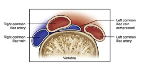
Very interesting and I'll wager this is what is happening in my case. (however it is my right common iliac vein that is the victim!)Cece wrote:I ran across this illustration and found it was helpful for visualizing May Thurner:
See the squished left iliac vein?
Sad to say that I went in March to have this looked at with IVUS. The doctor went in my left groin, turned the corner at my IVC junction, looked at it under x-ray, thought it looked fine and so he never looked at it using IVUS!!!
Man, was I pissed!!!
I am still considering another follow-up with our Brooklyn buddy next year because my insurance is switching. I know he'll look at it with IVUS but what a huge delay!!!
Cece, what was the source for this?
Thanks!
If you can't explain it simply, you don't understand it well enough. - Al Einstein
It's from this powerpoint presentation on IVUS in "iliocaval" disease:
www.fvs.org/2011Powerpoint/Sun_0925Almeida.pps
It's about halfway through. If I'm understanding that page it's on, it shows a majority presentation and a minority presentation. The minority presentation is in 22% of May Thurners cases and is how a patient would get a right proximal iliac compression. Let me know if you see it the same way.
How frustrating not to have it fully examined. Better luck next time....