IVUS

A forum to discuss Chronic Cerebrospinal Venous Insufficiency and its relationship to Multiple Sclerosis.
User avatar
drsclafani
Family Elder
Posts: 3182
Joined: Fri Mar 12, 2010 3:00 pm
Location: Brooklyn, New York
Contact:

Post by drsclafani »

CureOrBust wrote:
drsclafani wrote:
CureOrBust wrote:I understand this question may be a bit much to ask at this early stage, but roughly, or if at all, what percentage chance do you think would there be of someone not showing issues on a standard external Doppler and no issues in a venography, but IVUS would find something? from a purely diagnosis perspective.
Perhaps it is just me, but i find that the majority of azygous valvular abnormalities are not visible on venography but visible on IVUS.

But IVUS is not just about making a diagnosis. it is also about a treatment plan, balloon sizes, and assessment of the effects of angioplasty. It can also detect dissections after angioplasty that may go unrecognized without ivus.
I was obviously not clear enopugh, my question was if these issues were not visible under fluoroscope, would they present themselves as flow issue under a standard CCSVI doppler; errr... by standard I guess we would have to take Zamboni's measures...
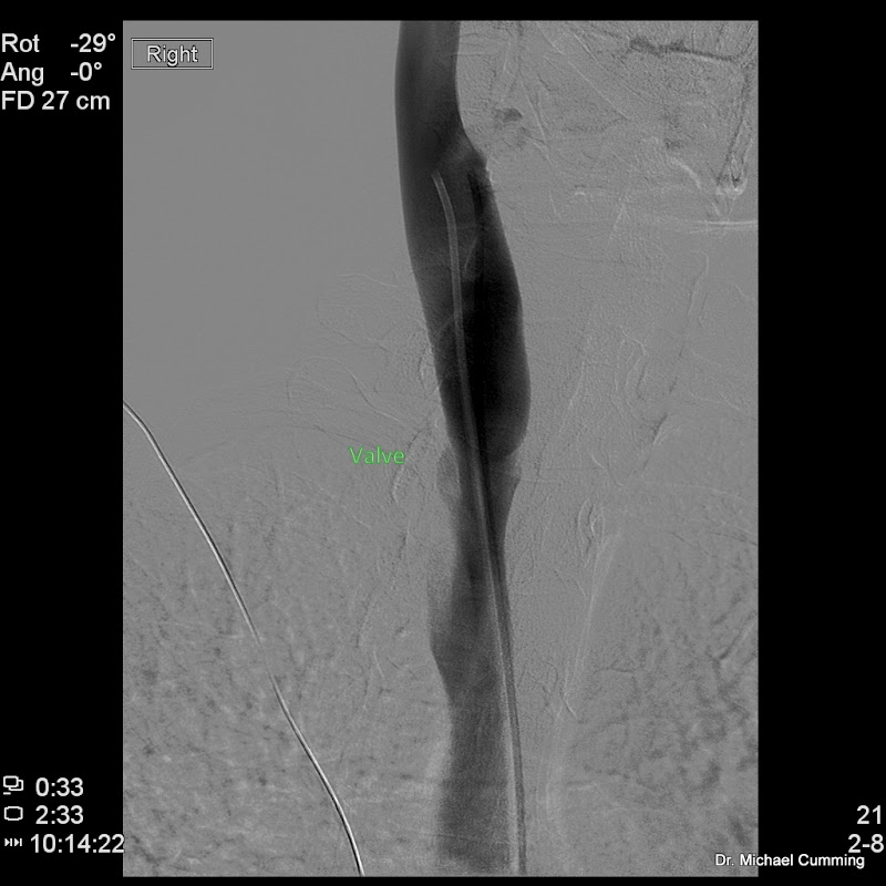
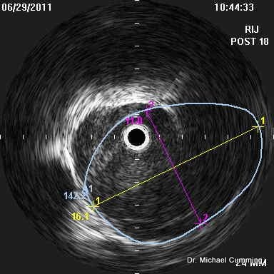
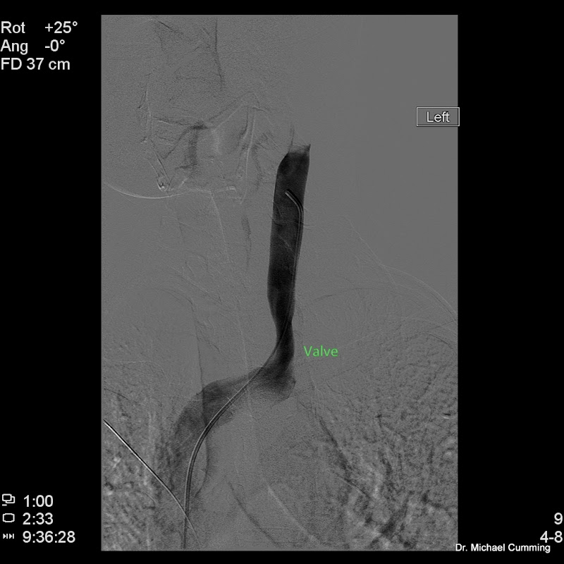
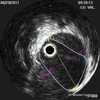
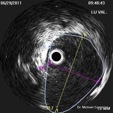
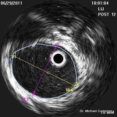
Firstly, in PwMS, surface B-mode and Doppler ultrasound almost always shows hemodynamic derangements when performed by the zamboni protocol and performed and interpreted by someone qualified to do so by training.

Secondly, these ultrasound abnormalities reflect problems with the global hemodynamics of the cerebrospinal venous circults

thirdly, they do not usually reflect some particular pathologic site of stenosis

Thus ultrasound is extremely sensitive to deranged hemodynamics but is n ot specific as to the veins that are problematic.

Ultrasound can rarely be used to pinpoint problems in the azygous vein.


Secondly
User avatar
CureOrBust
Family Elder
Posts: 3374
Joined: Wed Jul 27, 2005 2:00 pm
Location: Sydney, Australia

Post by CureOrBust »

Thanks for that. I already realised that the ultrasound would not give the exact "where", but you have now made it clear that "ultrasound is extremely sensitive to deranged hemodynamics". Now its a question of the sonographers knowledge.
User avatar
DrCumming
Family Elder
Posts: 209
Joined: Sat Dec 25, 2010 3:00 pm
Location: Minneapolis, MN
Contact:

Post by DrCumming »

Case 5: 50 yo female with RRMS

Images from both IJ's

Very classic findings. Bilateral IJ stenosis secondary to poorly opening valve leaflets. Excellent examples of using IVUS for pre and post ballooning.

Right:

This side is dominant.

Venogram prior to ballooning showing the stenosis at the level of the expected location of the valve plane.

Image

and IVUS better showing the valves and the size of the normal vein at the level of the valves. Knowing this allows better selection of balloon size.

Area at valves is 96 mm sq

Image

Vein at this level is 160 mm sq

Image

Initial ballooning was 16 mm. This was suboptimal on IVUS. Next ballooning was 18mm. Repeat IVUS shows improvement to 142 mm sq. Up for debate is should I have gone larger. Note in the top right corner, the valve leaflet is still not completely open. I do not have a good answer. The enemy of good is perfect so I elected to leave this alone. Its a substantial improvement from the original area of 96mm sq

Image

Post 18mm venogram looks good. You can see the one leaflet that is still not 100% open.

Image


Left:

This side is smaller then the right but not hypoplastic.

Venography shows no real narrowing. Yes, the vein does narrow as it enters the low neck. Trying to make this segment of vein look like the mid portion of the IJ will often result in occlusion or severe restenosis. See discussion below.

Image

But IVUS at the valve plane shows the incompletely opening valve leaflets with an area of 49 mm sq.

Image

The area of the jugular vein at this level is 96 mm sq.

Image

The area of the jugular vein just above the valve plane is 62 mm sq (Not shown).

So, this raises some interested questions about how to treat the left side.

1. Do we make the opening of the valves equal to the area of the jugular vein immediately distal?

2. Make the valves open to the size of the vein at the level of the valves? Can we do this without causing significant injury to the "normal" smaller jugular vein just distal to the valves?

3. Make the entire proximal segment of the jugular vein as big as the mid portion of the jugular vein?

Number 3 in my experience often results in occlusion or severe restenosis. I do not do this anymore.

So, we are left with 1 and 2. 1 is definitely going to be the less traumatic option (in terms of intimal injury). But may not result in optimal treatment benefit. Option 2 may work, but also may cause too much intimal injuryto the vein just distal. I don't have a good answer but I was requested to do the procedure to minimize risk. So I chose option 1.

Initial dilation to 10 mm did not improve the area. Dilation to 12 mm improved the area to 62 mm sq.

Image

And post venogram is not all that different (as expected).

Image


So, some very good images and examples of the complexity of the decision making behind these procedures and the many things we do not know. Yet.
User avatar
drsclafani
Family Elder
Posts: 3182
Joined: Fri Mar 12, 2010 3:00 pm
Location: Brooklyn, New York
Contact:

Post by drsclafani »

DrCumming wrote:Case 5: 50 yo female with RRMS

Images from both IJ's

Very classic findings. Bilateral IJ stenosis secondary to poorly opening valve leaflets. Excellent examples of using IVUS for pre and post ballooning.

Right:

This side is dominant.

Venogram prior to ballooning showing the stenosis at the level of the expected location of the valve plane.

Image

and IVUS better showing the valves and the size of the normal vein at the level of the valves. Knowing this allows better selection of balloon size.

Area at valves is 96 mm sq

Image

Vein at this level is 160 mm sq

Image

Initial ballooning was 16 mm. This was suboptimal on IVUS. Next ballooning was 18mm. Repeat IVUS shows improvement to 142 mm sq. Up for debate is should I have gone larger. Note in the top right corner, the valve leaflet is still not completely open. I do not have a good answer. The enemy of good is perfect so I elected to leave this alone. Its a substantial improvement from the original area of 96mm sq

Image

Post 18mm venogram looks good. You can see the one leaflet that is still not 100% open.

Image


Left:

This side is smaller then the right but not hypoplastic.

Venography shows no real narrowing. Yes, the vein does narrow as it enters the low neck. Trying to make this segment of vein look like the mid portion of the IJ will often result in occlusion or severe restenosis. See discussion below.

Image

But IVUS at the valve plane shows the incompletely opening valve leaflets with an area of 49 mm sq.

Image

The area of the jugular vein at this level is 96 mm sq.

Image

The area of the jugular vein just above the valve plane is 62 mm sq (Not shown).

So, this raises some interested questions about how to treat the left side.

1. Do we make the opening of the valves equal to the area of the jugular vein immediately distal?

2. Make the valves open to the size of the vein at the level of the valves? Can we do this without causing significant injury to the "normal" smaller jugular vein just distal to the valves?

3. Make the entire proximal segment of the jugular vein as big as the mid portion of the jugular vein?

Number 3 in my experience often results in occlusion or severe restenosis. I do not do this anymore.

So, we are left with 1 and 2. 1 is definitely going to be the less traumatic option (in terms of intimal injury). But may not result in optimal treatment benefit. Option 2 may work, but also may cause too much intimal injuryto the vein just distal. I don't have a good answer but I was requested to do the procedure to minimize risk. So I chose option 1.

Initial dilation to 10 mm did not improve the area. Dilation to 12 mm improved the area to 62 mm sq.

Image

And post venogram is not all that different (as expected).

Image


So, some very good images and examples of the complexity of the decision making behind these procedures and the many things we do not know. Yet.
Mike
good case and some provocative questions. I do not have the answers yet, at some point we should put our data together but it should only include good clinical and imaging followup.

i am thinking that the size of the contrast column is not the issue, the issue is whether the leaflets are pulled apart sufficiently to delay or prevent restenosis of the valve.

I agree that over dilation results in risks so i am more cautious but by using sequential dilatation i have been able to reduce risks. I still select balloons that are greater than the CSA of the vein in order to push the valve leaflets apart. I personally dont think that we will get the desired effect for sufficiently long if we dont So i continue to use the 50-100% increase above nominal diameter, starting at closer to 50% and then increasing balloon size if necessary to get the valve completely open.

i have had one thrombosis in the last 60 or so cases, plus two dissections that were unrelated to balloon size but to balloon rupture. Both have healed. This comes while i am more aggressive at early detection of thrombus.
User avatar
DrCumming
Family Elder
Posts: 209
Joined: Sat Dec 25, 2010 3:00 pm
Location: Minneapolis, MN
Contact:

Post by DrCumming »

drsclafani wrote:Mike
good case and some provocative questions. I do not have the answers yet, at some point we should put our data together but it should only include good clinical and imaging followup.

i am thinking that the size of the contrast column is not the issue, the issue is whether the leaflets are pulled apart sufficiently to delay or prevent restenosis of the valve.

I agree that over dilation results in risks so i am more cautious but by using sequential dilatation i have been able to reduce risks. I still select balloons that are greater than the CSA of the vein in order to push the valve leaflets apart. I personally dont think that we will get the desired effect for sufficiently long if we dont So i continue to use the 50-100% increase above nominal diameter, starting at closer to 50% and then increasing balloon size if necessary to get the valve completely open.

i have had one thrombosis in the last 60 or so cases, plus two dissections that were unrelated to balloon size but to balloon rupture. Both have healed. This comes while i am more aggressive at early detection of thrombus.
Hi Sal,

And yes, we need to put together our procedural data to best understand how to treat. There is much to be learned.

Next case is a patient with early restenosis - one side from under treating and the other from over treating....

Mike

Thanks.
User avatar
DrCumming
Family Elder
Posts: 209
Joined: Sat Dec 25, 2010 3:00 pm
Location: Minneapolis, MN
Contact:

Case 6:

Post by DrCumming »

Case 6: 49 yo male, RRMS

Will split into 2 posts.

First, the right side.

Preprocedure conventional US nicely shows the poorly opening valve leaflets.

Image

Image

and IVUS


Image

Note the fairly good agreement between in area between the conventional US and IVUS.

Venogram


Image

Notice the valvular stenosis cannot be seen. The valves are oriented in a coronal plane which makes seeing the stenosis very difficult on venograpgy. Only a lateral venogram could show the stenosis. I tried and couldn't get a good enough picture to show it.

So, we have a valve area of 92 mm sq, area at annulus (not shown) of 140 mm sq, and area at the incompletely opening valves of 186 mm sq.

Started with a 16 mm balloon (area is about 200 mm sq), about a 40% oversize relative to the annulus and about equivalent to the area at the valve stenosis. Repeat IVUS showed improvement (not shown) but residual stenosis.

Next, used an 18 mm balloon (area about 254 mm sq). This improved the area of the valves to 125 mm sq. Almost the same as the vein at the level of the annulus but not as large as the vein at the level of the valves.

Image

Good enough or not? (Sal, I know what you are going to say...)

Patient improved significantly for 2 weeks and then worsened back to baseline.

Repeat conventional US shows worsening of valve stenosis....

Image

and

Image


So, repeat venoplasty, this time using 20mm balloon being careful to try and NOT dilate the annulus, just the valves.

Here is the repeat IVUS


Image

and post 20 mm IVUS showing no valves

Image

and then repeat conventional US while on the procedure table show no identifiable valves....

Image


Nice case showing good correlation between conventional US and IVUS and a good example of under treatment. Will see if more aggressive venoplasty is more durable.

I will post the left side later. What is really interesting on the left side, is the patient had severe restenosis due to intimal hyperplasia (ie over treatment). Yet, we will see, that the venoplasty on this side was conservative as well. Confusing...
Cece
Family Elder
Posts: 9335
Joined: Mon Jan 04, 2010 3:00 pm
Contact:

Post by Cece »

I will post the left side later. What is really interesting on the left side, is the patient had severe restenosis due to intimal hyperplasia (ie over treatment). Yet, we will see, that the venoplasty on this side was conservative as well. Confusing...
What was the patient on for anticoagulation? Do you think an antiplatelet does anything to help against intimal hyperplasia? I am surprised (and saddened) to hear that the patient developed this even after conservative treatment.

Was it the left vein that developed the intimal hyperplasia? Have you noticed any more complications with the left vein as opposed to the right vein? (In many patients, the right vein is dominant or bigger.)

If the patient had improvements for two weeks and then worsened, how long did you wait before the second procedure? Should there be any concerns about having procedures too close together, can that aggregate any damage that might occur to the vein wall?

One thing I love is that this patient is receiving follow-up care. For the longest time, we heard of patients getting treated in far-off places and there was no opportunity for follow-up. Things are getting better for us.

Can't wait to see the rest of this case!
User avatar
HappyPoet
Family Elder
Posts: 1414
Joined: Thu Jul 09, 2009 2:00 pm
Contact:

Post by HappyPoet »

drcumming wrote:and then repeat conventional US while on the procedure table show no identifiable valves....
Do you use before and after conventional US as a standard part of your protocol for all your procedures?

Thank you!
User avatar
DrCumming
Family Elder
Posts: 209
Joined: Sat Dec 25, 2010 3:00 pm
Location: Minneapolis, MN
Contact:

Post by DrCumming »

HappyPoet wrote:
drcumming wrote:and then repeat conventional US while on the procedure table show no identifiable valves....
Do you use before and after conventional US as a standard part of your protocol for all your procedures?

Thank you!
Absolutely (except for the patients that travel - although many do come back). Its the only way to learn and modify our approach. I don't normally do it on the table. This time I had to make sure I couldn't see those #$#$&)% valves any more :)
User avatar
DrCumming
Family Elder
Posts: 209
Joined: Sat Dec 25, 2010 3:00 pm
Location: Minneapolis, MN
Contact:

Post by DrCumming »

Cece wrote:
I will post the left side later. What is really interesting on the left side, is the patient had severe restenosis due to intimal hyperplasia (ie over treatment). Yet, we will see, that the venoplasty on this side was conservative as well. Confusing...
What was the patient on for anticoagulation? Do you think an antiplatelet does anything to help against intimal hyperplasia? I am surprised (and saddened) to hear that the patient developed this even after conservative treatment.

Was it the left vein that developed the intimal hyperplasia? Have you noticed any more complications with the left vein as opposed to the right vein? (In many patients, the right vein is dominant or bigger.)

If the patient had improvements for two weeks and then worsened, how long did you wait before the second procedure? Should there be any concerns about having procedures too close together, can that aggregate any damage that might occur to the vein wall?

One thing I love is that this patient is receiving follow-up care. For the longest time, we heard of patients getting treated in far-off places and there was no opportunity for follow-up. Things are getting better for us.

Can't wait to see the rest of this case!
Not so much left or right but smaller vs larger. Small are hard to deal with. Although I have another example to post of occlusion at one month of the large dominant vein and a widely patent smaller (hypoplastic) vein.

Not sure any medication is going to help with this problem.

I keep meaning to do a restenosis thread... just not enough time. But here is one (crazy) thought. When we injury the vein wall with the balloon, we denude/tear the endothelium. The next layer, the media, is "sticky". When you are erect the jugular veins collapse. The sticky surfaces touch each other and stick together. I have seen this in some pig models that I worked on a long time ago. So, rather than an inclined bed post ballooning, maybe people should lay flat for a few days.... This would keep the IJ fully expanded while it is healing. Just a crazy guess. Short term restenosis (ie < 1 month) is not just simple intimal hyperplasia. There is more to it than that.
Cece
Family Elder
Posts: 9335
Joined: Mon Jan 04, 2010 3:00 pm
Contact:

Post by Cece »

This media, it's not sticky in a clotting sense. Just sticky....

I agree about the inclined bed, I kept mine flat for a month post-procedure in February.

I think there's logical support for encouraging people to lie flat as much as possible while it is healing, especially in the beginning. It's restrictive but if it helps, it helps. We've got a lifetime to enjoy the blood flow if the veins stay open or to regret it if they don't.

My crazy thought is, taking this further, if a patient is up and about while the veins are still healing, would it help to occasionally hold one's breath? The rationale would be that this would increase the blood in the jugulars and get those sticky walls away from each other, at least for the time being. I've seen how it looks on IVUS when the vein expands.
User avatar
DrCumming
Family Elder
Posts: 209
Joined: Sat Dec 25, 2010 3:00 pm
Location: Minneapolis, MN
Contact:

Post by DrCumming »

Follow up on Case #6

Did well for 1 week and then symptoms returned.

Here is the repeat US

Image

Right IJ valves are back and are fixed exactly as before.

Below is the US of the right IJ immediately after the last ballooning. Valves were not visible at all. Same with post procedure IVUS

Image

Sure would love to have a percutaneous valulotome.

2 tries at venoplastly. Any other options then a stent for round 3?
User avatar
ttucker
Getting to Know You...
Posts: 10
Joined: Sun Jul 10, 2011 2:00 pm

Post by ttucker »

While measuring CSA at various points in the vein, is it also possible to measure systolic and diastolic pressures at those points? With such measurements it may be possible to model the pressure distribution in the vein using computational fluid dynamics and to extrapolate that pressure distribution up into the area of the lesions. If that critical area is sufficiently hypertensive to cause disruption of the blood-brain barrier this could be a significant breakthrough in an understanding of the etiology of MS.
Dr. Trev Tucker
User avatar
MarkW
Family Elder
Posts: 1167
Joined: Thu Oct 19, 2006 2:00 pm
Location: Oxfordshire, England
Contact:

MS is multifactorial and CCSVI is a syndrome

Post by MarkW »

Hello Dr T,
Please keep clear of explaining the etiology of MS just because there is increased back pressure on the BBB. Its great that solving vein value issues increases venous flow and probably CSF clearance. I can knock down any MS etiology theory which relies solely on one cause, so please avoid this. The neuros argue that veins should not be treated because there is no proof that they are the cause of MS, they are wrong.
Prof Zamboni has stated MS is multifactorial and CCSVI is a syndrome, lets treat the syndrome and theorise later. Explain astrocyte death for me if you want to discuss details of MS etiology.

Kind regards,
Mark
Mark Walker - Oxfordshire, England. Retired Industrial Pharmacist. 24 years of study about MS.
CCSVI Comments:
http://www.telegraph.co.uk/news/health/8359854/MS-experts-in-Britain-have-to-open-their-minds.html
User avatar
ttucker
Getting to Know You...
Posts: 10
Joined: Sun Jul 10, 2011 2:00 pm

Post by ttucker »

Mark
I am a strong advocate of MS being a consequent of the perfect storm of multiple factors. I do try to bring this out in my Youtube and also in a paper I have had accepted for publication in one of the medical journals. Physics of fluid dynamics does not address other contributing factors like gender, genetics, diet, etc, but physics has been rather overlooked as a contributing factor.
Trev. Tucker
Post Reply

Return to “Chronic Cerebrospinal Venous Insufficiency (CCSVI)”