Cece, thanks for sharing and labeling your great images.
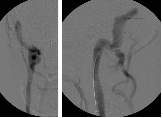
It looks like DrS brought the catheter into the Transverse Sinus (TS) just a bit before releasing the contrast dye. Very good news to learn your Sigmoid Sinuses (SSs) are properly connected to their respective IJVs with no SS stenoses. Very interesting to actually see an image of the vertebral veins (VVs) with dye going down them.
Images 1a, 1b (R-IJV, lateral): Is the vein to the right of the Sigmoid sinus the Right VV?
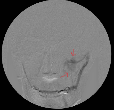
Image 3a (R-IJV, front): I think the blue arrow is the transverse dural sinus.
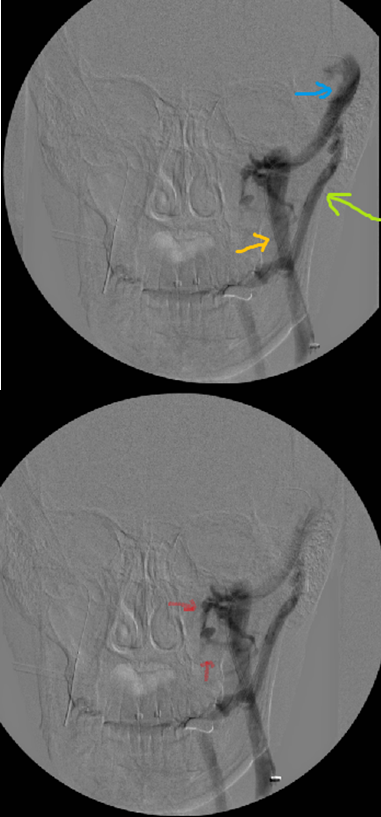
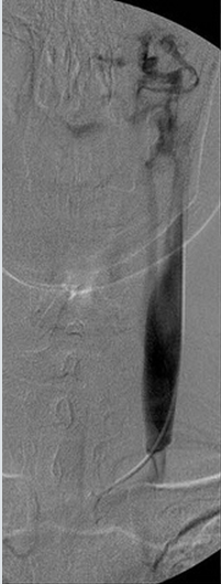
Images 2 and 3b (L-IJV, front): Any guess as to what the "squiggles" are? Vertebrals? Collaterals? Plexus veins?
Image 4 (L-IJV, lateral): Again (see Image 1a, 1b), is the vein to the right of the Sigmoid sinus the Left VV?
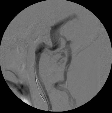
Do you have any post-valvuloplasty images showing if the VVs stopped "lighting up" with dye, or would that necessitate DrS having to go back into the SS to release more dye and use more radiation? Is it safe to believe that the VVs won't fill with dye anymore once the lower IJV valvuloplasty is completed?
~~~~~~~~~~
Cece, you set the bar very high with your searching abilities.
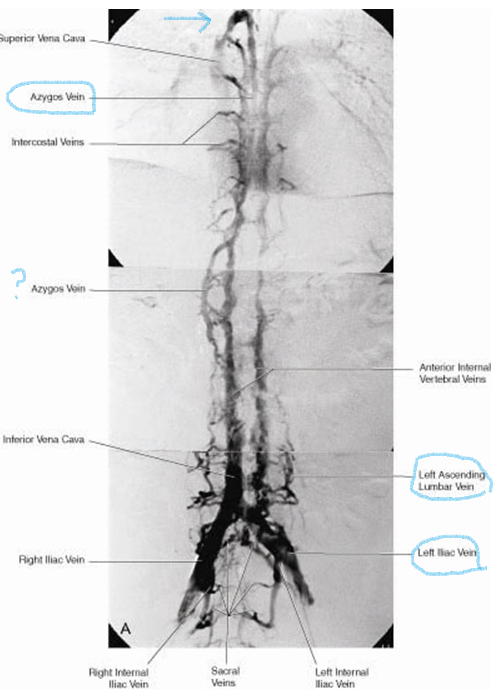
The composite image showing all those veins is fantastic. I was always unsure where exactly the lumbar veins are, and seeing them in this image clears up my confusion. I'm still unsure of where the renal veins are? I agree that the vein to the left of the Azygos is the Hemiazygos vein.
Here is a link to a great PowerPoint presentation that shows the dural sinuses and the bottom of the skull in all their glory (the d/l might be a bit slow). One can really see how vulnerable all the vessels and nerves are to shifts in the skull:
www.similima.com/ppt/anatomy/anat-cranial-fossa.ppt
Thanks, Cece! Great job!!


