Dorothy
Cece's turn
-
Kathryn333

- Family Member
- Posts: 45
- Joined: Sun Jul 25, 2010 2:00 pm
- Location: Toronto, Ontario
- Contact:
Re: Cece's turn
Hi, Cece. I'm so happy to read your latest update. Continue to heal. Congratulations. 
Dorothy
Dorothy
-
Cece
- Family Elder
- Posts: 9335
- Joined: Mon Jan 04, 2010 3:00 pm
- Contact:
Re: Cece's turn
So much support and well wishes here. Thank you.
I feel a little self-conscious talking about kickboxing here. I mention it because there are profound changes that show themselves when I exercise. (Sweating, balance, endurance, not getting ill from exercise.)
Cheer, from the beginning, how you described his MS resonated with my own. I had the similarities with having relapses brought on by altitude. (Hiking is good, hiking up high is not good.) I hope it lasts for me the way it has lasted for him!
-
Jugular

- Family Elder
- Posts: 375
- Joined: Mon Dec 21, 2009 3:00 pm
- Contact:
Re: Cece's turn
I hope you continue to improve, enjoy the color-drenched world, and have reason to sing
"Sha la la la la la la la la la la te da "
"Sha la la la la la la la la la la te da "
-
HappyPoet

- Family Elder
- Posts: 1414
- Joined: Thu Jul 09, 2009 2:00 pm
- Contact:
Re: Cece's turn
Kickboxing! Wow! You go girl! And don't let anything stop you!
-
Cece
- Family Elder
- Posts: 9335
- Joined: Mon Jan 04, 2010 3:00 pm
- Contact:
Re: Cece's turn
kodachrome, don't you know!Jugular wrote:I hope you continue to improve, enjoy the color-drenched world, and have reason to sing
"Sha la la la la la la la la la la te da "
-
Cece
- Family Elder
- Posts: 9335
- Joined: Mon Jan 04, 2010 3:00 pm
- Contact:
Re: Cece's turn
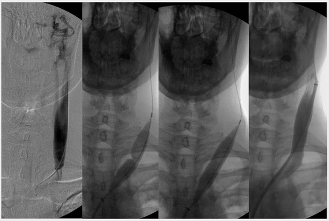
My left jugular, procedure #1:
-
Cece
- Family Elder
- Posts: 9335
- Joined: Mon Jan 04, 2010 3:00 pm
- Contact:
Re: Cece's turn
my left jugular, procedure #2, five months later:
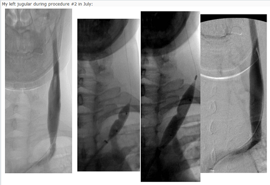
The end result looks equally good both times.
It has lasted without restenosing longer this time. I am still doing really well.
Even before procedure #2, in the first image of the second set, it is a more functional vein than it was before the first procedure.
In that first image of the second set, it could be restenosis due to scarring, but Dr. Sclafani thought it was restenosis due to residual valve material. Or restenosis due to collagen III abnormalities that made the valve less compliant? It's not clotting and it's not intimal hyperplasia. It could be considered natural narrowing before the jugular joins the subclavian but it was seen that the contrast just sat in the vein, in stasis, not going anywhere. And I had improvements upon treatment.
-
Cece
- Family Elder
- Posts: 9335
- Joined: Mon Jan 04, 2010 3:00 pm
- Contact:
Re: Cece's turn
I have not updated in a while but everything is still good. Twice I've had a few weeks or a month where colors don't seem as bright (in November and in January, if I'm recalling right) and then, bam, they're back, and I can't walk through a mall or a gym without the colors grabbing my attention and seeming all unnatural. It was especially odd in January to feel like the colors were lessened, because I had improvements at the start of January too. Still improving nearly a year out from the first procedure.
I also have a history of setbacks in the December/January timeframe. No setback this year.
I also have a history of setbacks in the December/January timeframe. No setback this year.
-
DougL

- Family Elder
- Posts: 384
- Joined: Mon Jul 11, 2011 2:00 pm
Re: Cece's turn
good to hear Cece
-
Cece
- Family Elder
- Posts: 9335
- Joined: Mon Jan 04, 2010 3:00 pm
- Contact:
Re: Cece's turn
This is a seriously long thread!
I wanted to update that I've arranged for an ultrasound check, which will be tomorrow with Dr. Cumming here in Minneapolis.
I've had a number of small symptoms that make me think I should get checked out. A week ago I had some foot drop issues recur for the first time since last May. I had a 100 degree fever, but I've had fevers between then and now and the foot drop didn't recur, so why this time. It's gone away now. I had a week when my left arm was having odd sensations, which I hadn't had since September. It never led to full-blown numbness and it went away. I had eye discomfort again, and man! That led to some curses. I have been taking my eyes for granted. Those are all symptoms that used to come and go, so it's hard to point to them too strongly, except that they had stayed very gone for a long time gone. On the more everyday front, instead of having dim colors all the time (pretreatment) or bright colors all the time (posttreatment) or dim colors all the time (restenosed) or bright colors all the time (after second treatment), I'm having bright colors some days and dim colors some days. Bright colors some weeks and dim colors some weeks. Crazy-making. I'm also finding myself waking up feeling ill some days, and it takes awhile to wear off, and that is familiar.
I'm still much better than I was pre-treatment. Back in January was when the change in colors really became apparent, and back in January was when I got marked improvements in bladder and diaphragm spasms. Those improvements have lasted. The cogfog improvements which came after the second treatment are as good as ever. Many other improvements too.
I am not sure what Dr. Cumming will see on ultrasound tomorrow and I am not sure what I would do depending on what he sees. We'll see what he advises.
-
DougL
- Family Elder
- Posts: 384
- Joined: Mon Jul 11, 2011 2:00 pm
Re: Cece's turn
you sound just like my partner.
post treatment there are good days and bad days.
pre treatment there were bad days and worse days (this was for about the last 5 years)
post treatment - she has days where her walking is 100% and others where she can barely stand on her own.
pre-treatment - her walking sucked (her words)
i wish you continued healing Cece
-
tiltawhirl
- Family Elder
- Posts: 126
- Joined: Sat Mar 17, 2012 7:47 pm
Re: Cece's turn
All the best in continued success Cece. You have no idea how valuable reading your posts was to me before making the decision to move forward.
My wife and I were and are grateful.
Tip of the hat, my dear. Keep us posted.
tilt
My wife and I were and are grateful.
Tip of the hat, my dear.
tilt
...and I for one, welcome our new Neurologist overlords!
My before and after CCSVI treatment video http://www.youtube.com/watch?v=RhosV4_DvWw
Visit my Lego Amusement Rides website http://www.brickshelf.com/cgi-bin/gallery.cgi?m=Bolliger
My before and after CCSVI treatment video http://www.youtube.com/watch?v=RhosV4_DvWw
Visit my Lego Amusement Rides website http://www.brickshelf.com/cgi-bin/gallery.cgi?m=Bolliger
-
newlywed4ever
- Family Elder
- Posts: 255
- Joined: Thu Apr 17, 2008 2:00 pm
- Location: Michigan
- Contact:
Re: Cece's turn
Cece, just want to let you know that I continue to follow posts here on TIMS. I don't often post but consider you and Dr Sclafani my mentors/teachers. Do keep us posted and, as always, I wish only the best for you.
-
erinc14
- Family Elder
- Posts: 599
- Joined: Sat Jun 12, 2010 2:00 pm
- Location: Montreal
Re: Cece's turn
best wishes for tomorrow !
-
cheerleader

- Family Elder
- Posts: 5361
- Joined: Mon Sep 10, 2007 2:00 pm
- Location: southern California
Re: Cece's turn
Cece--
Sending good thoughts. Don't discount an illness. Jeff thought he had restenosed last week, he had a return of fatigue, dizziness, pain, felt ill....turned out he has shingles. On an anti-viral, resting and feeling much better today. That fever may have been indicative of a flu or viral/bacterial infection. Hope you get some answers from Dr. Cumming, but if flow looks good, remember--not everything is MS/CCSVI-related.
Sending good thoughts. Don't discount an illness. Jeff thought he had restenosed last week, he had a return of fatigue, dizziness, pain, felt ill....turned out he has shingles. On an anti-viral, resting and feeling much better today. That fever may have been indicative of a flu or viral/bacterial infection. Hope you get some answers from Dr. Cumming, but if flow looks good, remember--not everything is MS/CCSVI-related.
Husband dx RRMS 3/07
dx dual jugular vein stenosis (CCSVI) 4/09
http://ccsviinms.blogspot.com
dx dual jugular vein stenosis (CCSVI) 4/09
http://ccsviinms.blogspot.com