Hi
As the MEDICOVER experience in CCSVI tretaments shows, approximately 4% of all cases cannot be treated by the simple "ballooning" procedure or by the implants of stents. The reasons are because the vein is pathologically too small (making "ballooning" ineffective) or because the vein is impeded in the natural funcion by a "strong" muscle nearby, which compresses the vein constantly, or because the stenosis is too long and the implant of stents would make the post-treatment thrombosis risk too high. In all these cases the treatment for CCSVI is NOT recommended by MEDICOVER because of the failure of the procedure to heal permanently too small veins or because of the predominant risk of any intervention. Contrary to the other Polish CCSVI treatment centers, MEDICOVER explains to any similar patient, why in these cases the procedure will NOT be performed: it does not bring any advantage or it is too risky for the patient.
There is no report of such refusals to intervene in the other Polish CCSVI clinics, which operate regardless of the advantages for the patient. This is clearly an attempt to receive the money for the operation knowing in advantage that the patient will not be healed at all or by exposing the patient to higher risks after the treatment. Are these other clinics serious? Please judge by yourself.
http://www.ccsvi-ms.pl/forum/viewtopic. ... c25978cbf6
Regards
Rici
CCSVI without success?: numbers in statistic do not lie
CCSVI without success?: numbers in statistic do not lie
Last edited by Rici on Wed Apr 06, 2011 12:49 am, edited 1 time in total.
-
soapdiva884

- Family Elder
- Posts: 167
- Joined: Mon Apr 05, 2010 2:00 pm
- Contact:
John only had one stenosis on one side per Dr. Sclafani. The aszygous was fine during both procedures. At the second procedure there was thought of some stenosis on the other side, so it was ballooned. But I am unclear how there can be a "typical CCSVI patient". This seems anything but typical with each and every patient. Just thinking outloud.....Cece wrote:The typical CCSVI patient has a blockage in both jugulars and one in the azygous. Even if one of the blockages is untreatable, there would still be two other blockages to be treated.
Boyfriend dx'd 6-6-06!!! RRMS............CCSVI procedure done on Nov. 13, 2010 and March 7, 2011 by Dr. Sclafani!
I've heard the number as low as 5% and as high as 95%! Would definitely be interested in what percentage you're seeing.DrCumming wrote:Actually, most patients do not have azygous lesions. Simka's presentation says only 5%. My experience using both venography and IVUS is a slighter high - need to look to give an exact number.
I double-checked Zamboni, here is what he saw:
http://fondazionehilarescere.org/pdf/12-2538-ANGY.pdfThe azygous vein was affected in 55/65 of cases; some of them had a membranaceous obstructions or a twisting, or less frequently, atresias and septums; in 12 cases, the azygous system showed stenosis at several points up to even atresia or agenesis of the lumbar plexuses 12/65 (Figure 2).
Is it at all possible that the variations in percentage of azygous issues (or other issues) found at different facilities depends in part on the mobility/disability of the people who go to different facilities? The various facilities may not share a similar cross-section of MS disability distribution. I am interested in seeing some of the results broken down across disability level, type of MS, duration of disease, previous treatments, etc., not just according to study or facility. Different variables would be intriguing. I hope I'm being clear -- I'm never sure! 
-
DrCumming

- Family Elder
- Posts: 209
- Joined: Sat Dec 25, 2010 3:00 pm
- Location: Minneapolis, MN
- Contact:
Definitely will depend on the types of patients. So, in Simka's experience, most patients are traveling to see him, and that might mean patients with less disabling MS are going there.bluesky63 wrote:Is it at all possible that the variations in percentage of azygous issues (or other issues) found at different facilities depends in part on the mobility/disability of the people who go to different facilities? The various facilities may not share a similar cross-section of MS disability distribution. I am interested in seeing some of the results broken down across disability level, type of MS, duration of disease, previous treatments, etc., not just according to study or facility. Different variables would be intriguing. I hope I'm being clear -- I'm never sure!
Part of the problem is defining what is a problem in the azygous and what is reported. If its a simple stenosis in the main azygous vein on venograpghy then that is fairly easy to quantify. Some of us (including myself) are also using IVUS and may see more abnormalities. The other component is the veins supplying the azygous. Some patients have these bizarre or unusual plexi. We don't have anyway to treat these. So, Simka maynot be reporting these findings but Zamboni does.
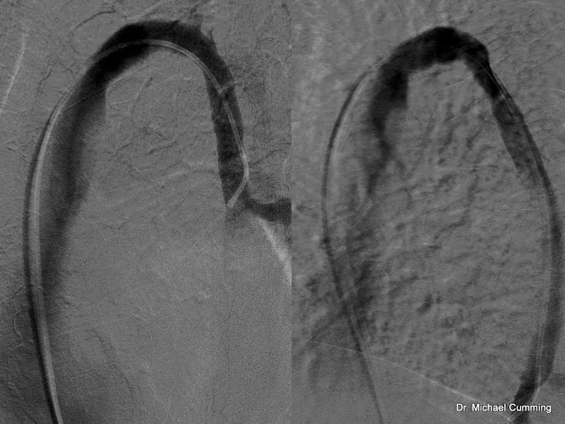
Here is an example below, left hand image was obtained at full inspiration, right image obtained at full expiration. The later shows a stenosis which is really isn't there. Makes sense when you think of the shape of the chest between the 2 states.
Last edited by DrCumming on Thu Mar 31, 2011 9:31 am, edited 1 time in total.
Your image did not work, I am interested. I thought end of expiration is exactly when it should be imaged?
(Intriguing points about how to define an azygous stenosis and about the travelling. Dr. Zamboni in particular would have had only Italian patients, compared to Dr. Simka's travelling patients.)
Here is EHC with the 95% azygos lesion figure:
http://www.essentialhealthclinic.com/we ... ions_.html
www.thisisms.com/ftopicp-140938.html#140938
On one hand, the worry as a patient is that an azygous stenosis will be missed; on the other hand, the worry is that it will be ballooned or stented needlessly.
Is twisting of the azygous a real phenomenon? I've wondered about that.
(Intriguing points about how to define an azygous stenosis and about the travelling. Dr. Zamboni in particular would have had only Italian patients, compared to Dr. Simka's travelling patients.)
Here is EHC with the 95% azygos lesion figure:
http://www.essentialhealthclinic.com/we ... ions_.html
Dr. Petrov from Bulgaria reports treating the azygous in 70% of patients:We have found over 95% of patients have azygous vein lesions which have required balloon angioplasty.
www.thisisms.com/ftopicp-140938.html#140938
On one hand, the worry as a patient is that an azygous stenosis will be missed; on the other hand, the worry is that it will be ballooned or stented needlessly.
Is twisting of the azygous a real phenomenon? I've wondered about that.
-
esta

- Family Elder
- Posts: 385
- Joined: Wed Nov 25, 2009 3:00 pm
- Location: Summerland. BC Canada
- Contact:
i am in a wheelchair and i went to katowice poland, as did many others and saw dr. simka. both times (for me), i had my azygos and even my iliac ckd - he said they were fine. buuuuuuuuuuuuttttttttttttt is my question to date, so i'm sitting back and waiting for more answers. ( btw i found handicap access way better overseas).
Last edited by esta on Thu Mar 31, 2011 9:41 am, edited 1 time in total.
PPMS. Liberated Katowice, Poland
06/05/10 angioplasty RJV-re-stenodsed
26/08/10 stent RJV
28/12/10 follow-up ultrasound intimal hyperplasia
06/05/10 angioplasty RJV-re-stenodsed
26/08/10 stent RJV
28/12/10 follow-up ultrasound intimal hyperplasia
-
Liberation

- Family Elder
- Posts: 339
- Joined: Fri Jan 07, 2011 3:00 pm
-
drsclafani

- Family Elder
- Posts: 3182
- Joined: Fri Mar 12, 2010 3:00 pm
- Location: Brooklyn, New York
- Contact:
mikeDrCumming wrote:fixed image link
expiration imaging was sal's idea. but i have seen this happen on several patients now. so, it may not be the best way to image the azygous.
in this case, I did expiration imaging, followed by ivus which showed no abnormality, and then did the imaging at full inspiration.
ideas are ideas. I throw out a lot of them. Not all pan out. Expiratory phase azygography didnt pan out. o, well.
i tend to first do end inspiration in the azygous for two reasons
1. less subtraction artefact for some reason
2. better distension of the azygous
Drs Zamboni and Galleoti often used the degree of reflux on the venogram to diagnose what they called subtle irregularities (webs). I have a great deal of difficulty seeing them, as do others. IVUS sometimes shows subtle sound reflections in the vein but i do not know what they represent.
IVUS definitely has shown abnormal valves in the arch that are not visible on Azygous venography tho.
The findings are much more difficult to analyze in the chest compared to the neck. Why? because there is constant motion during the chest venogram that we can generally avoid in the neck. You can stop breathing, but you cannot stop beating (the heart)