This Is MS Multiple Sclerosis Knowledge & Support Community
Welcome to This is MS, the leading forum for Multiple Sclerosis research and support. Join our friendly community of patients, caregivers, and researchers celebrating over 20 years of delivering hope through knowledge.
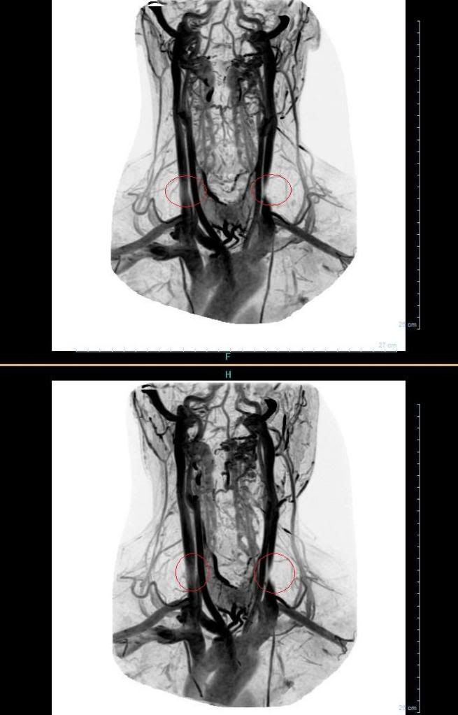
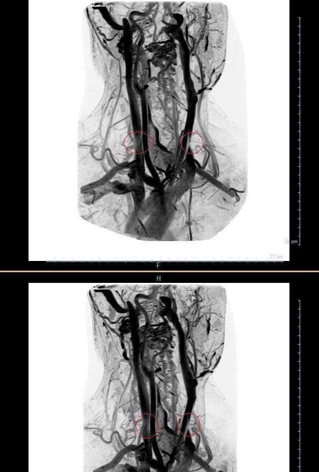
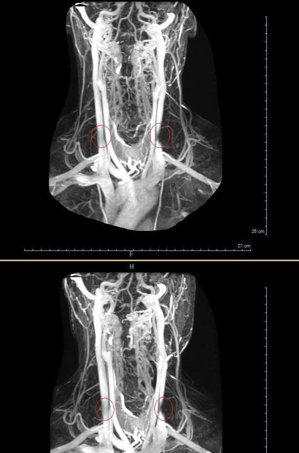
These MRV-pictures are the first one taken in Finland about CCSVI. I have been chatting with Blitzi today and weeks before. Getting to MRV wasn't easy.. but he succeeded to find a neuro who was interested. But I was a bit afraid that the lack of expertise makes this difficult - same things has happened elsewhere too I quess.
My wife's 3 yrs post video: http://www.youtube.com/watch?v=eLeqLps8XR8
Blitzi...
just wondering why the doc circled those areas of your jugulars in red if they are "normal"? They look a bit "different" to me, slightly hazy and pinched (but I'm not a doc) and in the final picture, there is a disconnect in the same area on the jugular on the right side, where the blood flow is broken off.
Are they willing to do the transcranial/neck dopplers as well, to check for reflux?
thanks for sharing the pics-
cheer
cheerleader wrote:Blitzi...
just wondering why the doc circled those areas of your jugulars in red if they are "normal"? They look a bit "different" to me, slightly hazy and pinched (but I'm not a doc) and in the final picture, there is a disconnect in the same area on the jugular on the right side, where the blood flow is broken off.
Are they willing to do the transcranial/neck dopplers as well, to check for reflux?
thanks for sharing the pics-
cheer
I Made those circles my self to point out what looks odd to me
But no, no transcranial doppler to me.... maybe if I again push my head trought the walls and make them do it.
That is only way in here finland now on.
These have very good quality compared to mine, looking more resolved and 3-dimensional. The former probably means higher magnetic field, the latter seems to be due to the fact that they're a composition of multiple images. Amazing. I second cheers thinking that there's a pinchig on one side. Did you do the circles yourself?
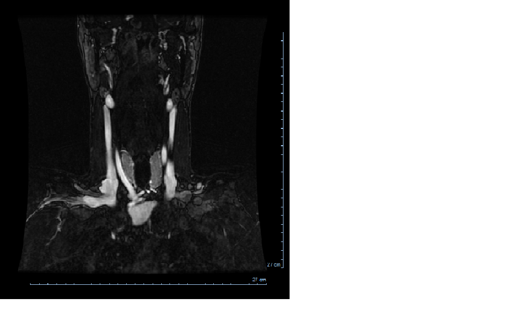
The pictures I saw of my jugulars that I thought showed the stenosis best were slices of the jugs as if you were stepping through the vein a slice at a time. There you could see how it dwindled down to nothing. Do you have any views like that? There were also views that showed the collateral veins very clearly. If I were you I'd send a copy of the CD to Dr Simka or Dr Dake.
It's true an MRI of the neck in the coronal plane right at the level of the pinching can show how "fat" and healthy the jugulars are at that level in a small slice at a time in the area where the blur is.
I am not a doctor either, but my neck MRI was the real convincer because it plainly showed the jugular pinched flat to the back.
I'm not offering medical advice, I am just a patient too! Talk to your doctor about what is best for you...
http://www.thisisms.com/ftopic-7318-0.html This is my regimen thread http://www.ccsvibook.com Read my book published by McFarland Health topics
Seems to my eyes tyou need to be "stented." My husband who took a look (a chiropractor) said that at the very least your subclavians need treatment. Did Dr. Dake see these pics? Daisyduck
These MRV-pictures are very new, taken yesterday and Blitzi hasn't even got the final statement from radiologist. We were wondering that is it possible that Sir Dake could take a look of Blitzi's mrvs?
My wife's 3 yrs post video: http://www.youtube.com/watch?v=eLeqLps8XR8
Ok, I now got final statement from radiologist, it says:
Shots taken from bottom of the skull to aorta, techincally very succefully images, veins have good contrast.
Sinussigmoidus and vena jugulars have normal fill, veins caliber are normal, except there is local narrow on down part of left vena jugularis internal which can be caused by something outside compression.
Cause by the compression might be sternocleidomastoid muscle.
Something is there?
Last edited by blitzi on Fri Oct 23, 2009 8:47 am, edited 1 time in total.
Looks like they are saying the sternocleidomastoid muscle might be compressing the base of the left internal jugular vein-and making it more narrow. It looks like that on all the pics, too, blitzi. Yes, there is something there. Any chance of getting dopplers or venography to check to blood flow and look for reflux? What's next?
cheer
Looks very close if not identical to the height of my narrowing. Hence I'm especially interested in the supposed cause. Can't find "sternogleinomastoideus muscle" on google. Did you come up with this possible cause, or one of your physicians? I find it interesting that the narrowing is at the same height on both sides (though more pronounced left in your case). This makes it less feasible that these are congenital defects, as why would they happen at the same height in two veins, but nowhere else?
Can I ask a random question: do you have or have you ever had a chronic sinus infection, making it difficult to breathe, or do you sniff a lot?
Don't know how Blitzi's story continues.. but I think it might be good idea to send these MRV:s to Finnish researches.. as some sort of evidence to CCSVI. I have been couple of times in contact with one of our head-researcher's and think that he is quite interested. These MRV's might give him more interest. But of course I have to talk with Blitzi about all this.
My wife's 3 yrs post video: http://www.youtube.com/watch?v=eLeqLps8XR8
Wikipedia entry: "[The sternocleidomastoid muscle] acts as an accessory muscle of inspiration, along with the scalene muscles of the neck."
Here we go. Since discussing my scans with Dake I tried to figure out what the muscles around my stenosis are used for and found that they contract whenever I sniff, apparently to aid holding up the windpipe. Now it so happens that I sniff a lot because of a chronic sinus infection I had for many years, and I have to use considerably more strength when inhaling through the nose than through the mouth. I seriously wonder whether this is what caused this narrowing in my case, and maybe in yours...?!
radeck wrote:Looks very close if not identical to the height of my narrowing. Hence I'm especially interested in the supposed cause. Can't find "sternogleinomastoideus muscle" on google. Did you come up with this possible cause, or one of your physicians? I find it interesting that the narrowing is at the same height on both sides (though more pronounced left in your case). This makes it less feasible that these are congenital defects, as why would they happen at the same height in two veins, but nowhere else?
Can I ask a random question: do you have or have you ever had a chronic sinus infection, making it difficult to breathe, or do you sniff a lot?
Muscle name was incorret, correct one is sternocleidomastoid I fix that on the post too.
That muscle conclusion is made by radiologist by himself.