Hello everyone.
Over the last few years I have had bouts of neurological symptoms, which I've dealt with as they happen, not linking them, some of which disappeared and others I am still left with. I had an Head MRI in October to exclude acoustic neuroma as a reason for my tinnitus and one sided deafness. This came back clear for tumours but the radiologist highlighted some periventricular hyperintensities which were too many to be age related and suggested demyelination and I was sent to a neurologist who said that the patches were not demyelination and the cause was likely to be vascular. He then asked about my history of high blood pressure, migraines, diabetes, high cholesterol as these would be the causes. I have never had any of these, nor any of my family members. He then said he'd do another MRI with contrast but signing me back to my GP as he expected it to come back normal. The results of the MRI are back and I would be grateful if you could shed some light on what they could possibly mean please:
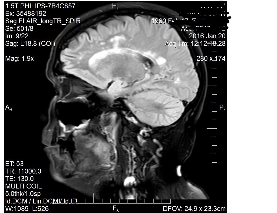
"There is again multifocal ill-defined and partially confluent T2/FLAIR hyperintensity in the deep white matter of both cerebral hemispheres. Some of this lies in a periventricular location adjacent to the occipital horns of both lateral ventricles. None of the lesions exhibit enhancement. No signal abnormality is seen within the midbrain, brainstem or cerebellum. A tiny focus is seen within the body of the corpus callosum. The distribution and extent has slightly progressed since 2011 (I had a brain scan done then for trigeminal neuralgia), but is unchanged since October 2015. It remains suspicious for demyelination. No other intracranial pathology is identified. Normal flow-voids are present within the major intracranial arteries and dural venous sinuses. The ventricular system itself has a normal configuration and the cerebellar tonsils are normally positioned."
I've now received a follow up letter from the neurologist:
I am pleased to say that your MRi scan is unchanged in comparison to October of last year, and in addition, there is no suggestion of abnormal activity when using contrast. This was what we had hoped to find. Overall I do not think there is anything significantly concerning here, and nothing that would indicate any specific additional treatment is required.
Yours Sincerely
Blah Blah Blah.
Don't know what to think or do now? He obviously thinks it's not demyelination despite three radiologists suggesting it.
New to the site - MRI results

Re: New to the site - MRI results
I think you are correct in suspecting possible early stage MS.
I base this up on the locations of the " T2/FLAIR hyperintensity" ...... periventricular location and corpus callosum. See the below link. The fact that there are no enhancing areas is very good. One key thing is there must be some progression to have a diagnosis of MS. You could have a one time event when you had a viral infection of some kind. I have no medical training but I have had MS since 1995.
The below link seems to be a good simple summary.
http://www.radiologyassistant.nl/en/p45 ... rosis.html
jackD
Re: New to the site - MRI results
Thank you for your reply JackD,
The first brain MRI that I had was for Trigeminal Neuralgia in 2011. I had a MRI in October 2015 to search for acoustic neuroma as I presented with one sided hearing loss, tinnitus and hyperacusis. I was then told about the radiologist's concerns about demyelination and I queried why hadn't it been found in the previous scan (2011) to which I was told that the radiologist had reported demyelination on that scan as well.
My neurologist seems to be of the opinion that, as the latest scan with contrast didn't show any activity, then there is nothing wrong? Does it always show when you have contrast? I have spoken to my GP about it and he said that he expects to have the same amount/distribution of lesions? when he's 70 - I wouldn't have minded that answer but as I told him (first time seeing him as he's new to the practice) I am a decade and a half away from that age, thank you very much!
Now I don't know what to do? Should I push for a second opinion? I did ask the neurologist if he was going to order an MRI of my spine but he refused. Should/can I push for a spinal MRI? Would that show anything new? The neurologist mentioned Lumbar Puncture? but just as quick dismissed it. The neurology appointment was me walking to the wall and turning (very unsteady), sitting on side of bed, finger to his finger, him testing my strength on my left side (right side too painful due to wrist fusion) then lying on bed after I took one shoe and sock off, and he asked if I could feel him on my foot and pulled my leg about a bit. He didn't bother with the left side, only the right side. The appointment was over so quickly that I ended up leaving him with a list of all the neurological things that have happened to be over the last few years. I doubt he even bothered looking at them to be honest. I just felt it was a waste of time.
-
lyndacarol

- Family Elder
- Posts: 3394
- Joined: Thu Dec 22, 2005 3:00 pm
- Contact:
Re: New to the site - MRI results
Welcome to ThisIsMS, Purrfik.Purrfik wrote:Over the last few years I have had bouts of neurological symptoms, which I've dealt with as they happen, not linking them, some of which disappeared and others I am still left with. I had an Head MRI in October to exclude acoustic neuroma as a reason for my tinnitus and one sided deafness. This came back clear for tumours but the radiologist highlighted some periventricular hyperintensities which were too many to be age related and suggested demyelination and I was sent to a neurologist who said that the patches were not demyelination and the cause was likely to be vascular. He then asked about my history of high blood pressure, migraines, diabetes, high cholesterol as these would be the causes. I have never had any of these, nor any of my family members. He then said he'd do another MRI with contrast but signing me back to my GP as he expected it to come back normal. The results of the MRI are back and I would be grateful if you could shed some light on what they could possibly mean please:
"There is again multifocal ill-defined and partially confluent T2/FLAIR hyperintensity in the deep white matter of both cerebral hemispheres. Some of this lies in a periventricular location adjacent to the occipital horns of both lateral ventricles. None of the lesions exhibit enhancement. No signal abnormality is seen within the midbrain, brainstem or cerebellum. A tiny focus is seen within the body of the corpus callosum. The distribution and extent has slightly progressed since 2011 (I had a brain scan done then for trigeminal neuralgia), but is unchanged since October 2015. It remains suspicious for demyelination. No other intracranial pathology is identified. Normal flow-voids are present within the major intracranial arteries and dural venous sinuses. The ventricular system itself has a normal configuration and the cerebellar tonsils are normally positioned."
I've now received a follow up letter from the neurologist:
I am pleased to say that your MRi scan is unchanged in comparison to October of last year, and in addition, there is no suggestion of abnormal activity when using contrast. This was what we had hoped to find. Overall I do not think there is anything significantly concerning here, and nothing that would indicate any specific additional treatment is required.
Yours Sincerely
Blah Blah Blah.
Don't know what to think or do now? He obviously thinks it's not demyelination despite three radiologists suggesting it.
I have no expertise in MRIs, but I do know that lesions in the brain are not specific to MS; lesions are common to MANY conditions.
Since neurological symptoms can be caused by vitamin D deficiency, which is VERY common today, I urge you to ask your GP for the vitamin D blood test. This is the 25-hydroxy D test. It is inexpensive (about $50).
Re: New to the site - MRI results
Thank you for the welcome LyndaCarol,
I had my Vitamin D levels checked earlier last year as part of a general check up (I have bone issues) and they were all within normal parameters. I do realise that MS is not the only cause of brain lesions and I am certainly not saying I do have it. Only that I would like to get to the reason for them in my brain. Whether that is MS or some other disease/condition etc. I have never lumped all my neurological conditions together ( unlike my daughter, who has said for years that I have MS) but have taken them one by one and dealt with them and carried on with life, albeit a bit slower each time.
I had my Vitamin D levels checked earlier last year as part of a general check up (I have bone issues) and they were all within normal parameters. I do realise that MS is not the only cause of brain lesions and I am certainly not saying I do have it. Only that I would like to get to the reason for them in my brain. Whether that is MS or some other disease/condition etc. I have never lumped all my neurological conditions together ( unlike my daughter, who has said for years that I have MS) but have taken them one by one and dealt with them and carried on with life, albeit a bit slower each time.
-
lyndacarol
- Family Elder
- Posts: 3394
- Joined: Thu Dec 22, 2005 3:00 pm
- Contact:
Re: New to the site - MRI results
I am curious. What were your vitamin D test result numbers last year? It is important to have the actual numbers. (The lab-established reference ranges for many tests are set notoriously low.)Purrfik wrote:I had my Vitamin D levels checked earlier last year as part of a general check up (I have bone issues) and they were all within normal parameters. I do realise that MS is not the only cause of brain lesions and I am certainly not saying I do have it. Only that I would like to get to the reason for them in my brain. Whether that is MS or some other disease/condition etc. I have never lumped all my neurological conditions together ( unlike my daughter, who has said for years that I have MS) but have taken them one by one and dealt with them and carried on with life, albeit a bit slower each time.
With bone issues, you are probably monitored on a regular basis. Some experts say that vitamin D should be tested in people like blood pressure, glucose, and cholesterol.
Re: New to the site - MRI results
Lynda, I was never given any numbers, just told that they all came back within normal parameters?
-
lyndacarol
- Family Elder
- Posts: 3394
- Joined: Thu Dec 22, 2005 3:00 pm
- Contact:
Re: New to the site - MRI results
In my opinion, it would be worthwhile to call your doctor's office and request a physical copy of those test results.Purrfik wrote:Lynda, I was never given any numbers, just told that they all came back within normal parameters?
"Normal" is not adequate. Normal is not the same as optimal.
Re: New to the site - MRI results
Thank you lyndacarol, I will do that.

Re: New to the site - MRI results
A deficiency in vitamin B12 can also cause lesions on MRI, including periventricular lesions.lyndacarol wrote:I have no expertise in MRIs, but I do know that lesions in the brain are not specific to MS; lesions are common to MANY conditions.
Since neurological symptoms can be caused by vitamin D deficiency, which is VERY common today, I urge you to ask your GP for the vitamin D blood test. This is the 25-hydroxy D test. It is inexpensive (about $50).
Please see the references in the following topic.
http://www.thisisms.com/forum/natural-a ... 24857.html
Purrfik, do you know if you've been tested for B12 deficiency?
Re: New to the site - MRI results
Hi NHE,
I do not know if I've been tested for B12 deficiency so I'll get hold of my GP and ask - normally a 6 week wait for routine appointments though so could take a while
I do not know if I've been tested for B12 deficiency so I'll get hold of my GP and ask - normally a 6 week wait for routine appointments though so could take a while
-
1eye

- Family Elder
- Posts: 3780
- Joined: Wed Mar 17, 2010 3:00 pm
- Location: Kanata, Ontario, Canada
- Contact:
Re: New to the site - MRI results
If I were you I wouldn't worry about it. Hasn't changed in a year, no enhancement (that means no leak across the blood-brain barrier). Keep living, eat well, exercise a lot. Walk a lot. Have a good life.
This unit of entertainment not brought to you by FREMULON.
Not a doctor.
"I'm still here, how 'bout that? I may have lost my lunchbox, but I'm still here." John Cowan Hartford (December 30, 1937 – June 4, 2001)
Not a doctor.
"I'm still here, how 'bout that? I may have lost my lunchbox, but I'm still here." John Cowan Hartford (December 30, 1937 – June 4, 2001)